Do you want to buy antibiotics online without prescription? https://buyantibiotics24h.net/ - This is pharmacy online for you!
Microsoft word - kt-14291.doc
K-ASSAY For the quantitative determination of E2 in serum, plasma and other biological fluids For Research Use Only. Not for use in diagnostic procedures.
Page 1 of 7 K-ASSAY Product Information Cat. No. KT-14291 INTENDED USE The kit is a competitive inhibition enzyme immunoassay technique for the in vitro quantitative measurement of E2 in serum, plasma and other biological fluids. For research use only. Not for use in diagnostic procedures. COMPONENTS
Reagents Quantity
Pre-coated, ready to use 96-well strip plate
MATERIALS REQUIRED BUT NOT SUPPLIED 1. Microplate reader with 450 ± 10 nm filter. 2. Precision single and multi-channel pipettes and disposable tips. 3. Eppendorf Tubes for diluting samples. 4. De-ionized or distilled water. 5. Absorbent paper for blotting the microtiter plate. 6. Container for Wash Solution. STORAGE All the reagents should be kept according to the labels on vials. The Calibrator, Detection Reagent A, Detection Reagent B and the 96-well strip plate should be stored at -20°C upon being received. The unused strips should be kept in a sealed bag with the desiccant provided to minimize exposure to damp air. Opened test kits will remain stable until the expiration date shown, provided it is stored as prescribed above. PRINCIPLE This assay employs the competitive inhibition enzyme immunoassay technique. A monoclonal antibody specific for E2 has been pre-coated onto a microplate. A competitive inhibition reaction is launched between biotin labeled E2 and unlabeled E2 (Calibrators or samples) with the pre-coated antibody specific for E2. After incubation the unbound conjugate is washed off. Next, avidin conjugated to
Page 2 of 7 K-ASSAY
Horseradish Peroxidase (HRP) is added to each microplate well and incubated. The amount of bound HRP conjugate is reverse proportional to the concentration of E2 in the sample. After addition of the substrate solution, the intensity of color developed is reverse proportional to the concentration of E2 in the sample. SAMPLE COLLECTION AND STORAGE Serum Use a serum separator tube and allow samples to clot for two hours at room temperature or overnight at 4°C before centrifugation for 20 minutes at approximately 1,000 x g. Assay freshly prepared serum immediately or store samples in aliquot at -20°C or -80°C for later use. Avoid repeated freeze/thaw cycles. Plasma Collect plasma using EDTA or heparin as an anticoagulant. Centrifuge samples for 15 minutes at 1,000 x g at 4°C within 30 minutes of collection. Remove plasma and assay immediately or store samples in aliquot at -20°C or -80°C for later use. Avoid repeated freeze/thaw cycles. Other biological fluids Centrifuge samples for 20 minutes at 1,000 x g. Remove particulates and assay immediately or store samples in aliquot at -20°C or -80°C for later use. Avoid repeated freeze/thaw cycles. Note: 1. Samples to be used within 5 days may be stored at 4°C, otherwise samples must be stored at -20°C
(≤1 month) or -80°C (≤2 months) to avoid loss of bioactivity and contamination.
2. When performing the assay slowly bring samples to room temperature. 3. Sample hemolysis will influence the result, so hemolytic specimen can not be detected. REAGENT PREPARATION
Bring all kit components and samples to room temperature (18-25°C) before use.
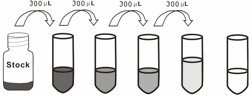
Calibrator Reconstitute the Calibrator with 0.5 mL of Calibrator Diluent, kept for 10 minutes at room temperature, shake gently (not to foam). The concentration of the calibrator in the stock solution is 1,000 pg/mL. Please prepare 5 tubes containing 0.6 mL Calibrator Diluent and produce a triple dilution series according to the picture shown below. Mix each tube thoroughly before the next transfer. Set up 5 points of diluted calibrator such as 1,000 pg/mL, 333.33 pg/mL, 111.11 pg/mL, 37.04 pg/mL, 12.35 pg/mL, and the last EP tubes with Calibrator Diluent is the blank as 0 pg/mL. 1,000 333.33 111.11 37.04 12.35 0
Page 3 of 7 K-ASSAY Assay Diluent A and B Dilute 6 mL of Assay Diluent A or B Concentrate (2X) with 6 mL of de-ionized or distilled water to prepare 12 mL of Assay Diluent A or B. The prepared working dilution can’t be frozen. Detection Reagent A and B Briefly spin or centrifuge the stock Detection Reagent A and Detection Reagent B before use. Dilute to the working concentration with working Assay Diluent A or B, respectively (1:100). Wash Solution Dilute 20 mL of Wash Solution Concentrate (30X) with 580 mL of de-ionized or distilled water to prepare 600 mL of Wash Solution (1X). TMB substrate Aspirate the needed dosage of the solution with sterilized tips and do not dump the residual solution into the vial again. Note: 1. Prepare calibrator within 15 minutes before assay. Pease do not dissolve the reagents at 37°C
2. Making serial dilution in the wells directly is not permitted. 3. Detection Reagent A and B are sticky solutions, therefore, slowly pipette them to reduce the volume
4. Please carefully reconstitute Calibrators or working Detection Reagent A and B according to the
instruction, and avoid foaming and mix gently until the crystals have completely dissolved. To minimize imprecision caused by pipetting, use small volumes and ensure that pipettors are calibrated. It is recommended to suck more than 10 µL for once pipetting.
5. The reconstituted Calibrators, Detection Reagent A and Detection Reagent B can be used only
6. If crystals have formed in the Wash Solution concentrate (30X), warm to room temperature and mix
gently until the crystals have completely dissolved.
7. Contaminated water or container for reagent preparation will influence the detection result. SAMPLE PREPARATION 1. Kamiya Biomedical Company is only responsible for the kit itself, but not for the samples consumed
during the assay. The user should calculate the possible amount of the samples used in the whole test. Please reserve sufficient samples in advance.
2. Please predict the concentration before assaying. If values for these are not within the range of the
calibration curve, users must determine the optimal sample dilutions for their particular experiments.
3. If the samples are not indicated in the manual, a preliminary experiment to determine the validity of
4. Tissue or cell extraction samples prepared by chemical lysis buffer may cause unexpected ELISA
results due to the impacts of certain chemicals.
5. Owing to the possibility of mismatching between antigen from other resource and antibody used in
our kits (e.g., antibody targets conformational epitope rather than linear epitope), some native or recombinant proteins from other manufacturers may not be recognized by our products.
6. Influenced by the factors including cell viability, cell number and also sampling time, samples from
cell culture supernatant may not be detected by the kit.
7. Fresh samples without long time storage is recommended for the test. Otherwise, protein degradation
and denaturalization may occur in those samples and finally lead to wrong results.
Page 4 of 7 K-ASSAY
ASSAY PROCEDURE
1. Determine wells for diluted calibrator, blank and sample. Prepare 5 wells for calibrator points, 1 well
for blank. Add 50 µL each of dilutions of calibrator (read Reagent Preparation), blank and samples into the appropriate wells, respectively. And then add 50 µL of Detection Reagent A to each well immediately. Shake the plate gently (using a microplate shaker is recommended). Cover with a Plate sealer. Incubate for 1 hour at 37°C. Detection Reagent A may appear cloudy. Warm to room temperature and mix gently until solution appears uniform.
2. Aspirate the solution and wash with 350 µL of 1X Wash Solution to each well using a squirt bottle,
multi-channel pipette, manifold dispenser or autowasher, and let it sit for 1~2 minutes. Remove the remaining liquid from all wells completely by snapping the plate onto absorbent paper. Repeat 3 times. After the last wash, remove any remaining Wash Buffer by aspirating or decanting. Invert the plate and blot it against absorbent paper.
3. Add 100 µL of Detection Reagent B working solution to each well. Incubate for 30 minutes at 37°C
after covering it with the Plate sealer.
4. Repeat the aspiration/wash process for total 5 times as conducted in step 2. 5. Add 90 µL of Substrate Solution to each well. Cover with a new Plate sealer. Incubate for 15 - 25
minutes at 37°C (Don’t exceed 30 minutes). Protect from light. The liquid will turn blue by the addition of Substrate Solution.
6. Add 50 µL of Stop Solution to each well. The liquid will turn yellow by the addition of Stop solution.
Mix the liquid by tapping the side of the plate. If color change does not appear uniform, gently tap the plate to ensure thorough mixing.
7. Remove any drop of water and fingerprint on the bottom of the plate and confirm there is no bubble on
the surface of the liquid. Then, run the microplate reader and conduct measurement at 450 nm immediately.
Note: 1. Assay preparation: Keep appropriate numbers of strips for 1 experiment and remove extra strips
from microtiter plate. Removed strips should be resealed and stored at -20°C until the kits expiration date.
2. Samples or reagents addition: Please use the freshly prepared Calibrator. Please carefully add
samples to wells and mix gently to avoid foaming. Do not touch the well wall as possible. For each step in the procedure, total dispensing time for addition of reagents or samples to the assay plate should not exceed 10 minutes. This will ensure equal elapsed time for each pipetting step, without interruption. Duplication of all calibrators and specimens, although not required, is recommended. To avoid cross-contamination, change pipette tips between additions of each calibrator level, between sample additions, and between reagent additions. Also, use separate reservoirs for each reagent.
3. Incubation: To ensure accurate results, proper adhesion of plate sealers during incubation steps is
necessary. Do not allow wells to sit uncovered for extended periods between incubation steps. Once reagents have been added to the well strips, DO NOT let the strips DRY at any time during the assay. Incubation time and temperature must be observed.
4. Washing: The wash procedure is critical. Complete removal of liquid at each step is essential to good
performance. After the last wash, remove any remaining Wash Solution by aspirating or decanting and remove any drop of water and fingerprint on the bottom of the plate. Insufficient washing will result in poor precision and falsely elevated absorbance reading.
5. Controlling of reaction time: Observe the change of color after adding TMB Substrate (e.g.
observation once every 10 minutes), if the color is too deep, add Stop Solution in advance to avoid excessively strong reaction which will result in inaccurate absorbance reading.
6. TMB Substrate is easily contaminated. Please protect it from light. 7. The environment humidity which is less than 60% might have some effects on the final performance,
therefore, a humidifier is recommended to be used at that condition.
Page 5 of 7 K-ASSAY CALCULATION OF RESULTS This assay employs the competitive inhibition enzyme immunoassay technique, so there is an inverse correlation between E2 concentration in the sample and the assay signal intensity.
Average the duplicate readings for each calibrator, control, and samples. Create a calibration curve on log-log or semi-log graph paper, with the log of E2 concentration on the y-axis and absorbance on the x-axis. Draw the best fit straight line through the calibrator points and it can be determined by regression analysis. Using some plot software is also recommended. If samples have been diluted, the concentration read from the calibration curve must be multiplied by the dilution factor.
PERFORMANCE Detection Range
The calibration curve concentrations used for the ELISA’s were 1,000 pg/mL, 333.33 pg/mL, 111.11 pg/mL, 37.04 pg/mL, 12.35 pg/mL. Sensitivity
The minimum detectable dose of E2 is typically less than 4.38 pg/mL. The sensitivity of this assay, or Lower Limit of Detection (LLD) was defined as the lowest protein concentration that could be differentiated from zero. It was determined the mean O.D. Value of 20 replicates of the zero calibrator plus three standard deviations. Specificity
This assay has high sensitivity and excellent specificity for detection of E2. No significant cross-reactivity or interference between E2 and analogues was observed.
Note: Limited by current skills and knowledge, it is impossible for us to complete the cross-reactivity detection between E2 and all the analogues, therefore, cross reaction may still exist.
ASSAY PROCEDURE SUMMARY 1. Prepare all reagents, samples and calibrators; 2. Add 50 µL calibrator or sample to each well. And then add 50 µL prepared Detection Reagent A
immediately. Shake and mix. Incubate 1 hour at 37°C;
3. Aspirate and wash 3 times; 4. Add 100 µL prepared Detection Reagent B. Incubate 30 minutes at 37°C; 5. Aspirate and wash 5 times; 6. Add 90 µL Substrate Solution. Incubate 15-25 minutes at 37°C; 7. Add 50 µL Stop Solution. Read at 450 nm immediately.
IMPORTANT NOTES 1. The final experimental results will be closely related to operation skills of the end users and the
experimental environments. Please make sure that sufficient samples are available.
2. Kits from different batches may be a little different in detection range, sensitivity and color developing
time. Please perform the experiment exactly according to the instruction attached in kit while electronic ones from our website (www.k-assay.com) is only for information.
3. Do not mix or substitute reagents from one kit lot to another. Use only the reagents supplied by
4. Protect all reagents from strong light during storage and incubation. All the bottle caps of reagents
should be covered tightly to prevent the evaporation and contamination of microorganism.
5. There may be some foggy substance in the wells when the plate is opened at the first time. It will not
have any effect on the final assay results. Do not remove microtiter plate from the storage bag until needed.
Page 6 of 7 K-ASSAY
6. Wrong operations during the reagents preparation and loading, as well as incorrect parameter setting
for the plate reader may lead to incorrect results. A microplate plate reader with a bandwidth of 10 nm or less and an optical density range of 0-3 O.D. or greater at 450 ± 10 nm wavelength is acceptable for use in absorbance measurement. Please read the instruction carefully and adjust the instrument prior to the experiment.
7. Even the same operator might get different results in two separate experiments. In order to get better
reproducible results, the operation of every step in the assay should be controlled. Furthermore, a preliminary experiment before assay for each batch is recommended.
8. Each kit has been strictly passed Q.C. test. However, results from end users might be inconsistent
with our in-house data due to some unexpected transportation conditions or different lab equipments. Intra-assay variance among kits from different batches might arise from above factors, too.
9. Kits from different manufacturers for the same item might produce different results, since we haven’t
compared our products with other manufacturers.
10. The Stop Solution suggested for use with this kit is an acid solution. Wear eye, hand, face, and
clothing protection when using this material.
FOR RESEARCH USE ONLY. Not for use in diagnostic procedures. www.k-assay.com
Page 7 of 7
UN DIA DE FIESTAS PERSONAJES POR ORDEN DE APARICIÓN l Carmen ( mujer de Paco) l Félix ( comisión de fiestas ) l Pepe ( comisión de fiestas ) l Paco ( albañil ) l Andrés ( Veterinario ) l Mariano ( alguacil ) l Eusebio ( pastor ) l Ana ( mujer de Veterinario ) l Raquel ( mujer de Alguacil) l Jugador de frontón Miguel Ángel l Jugador de frontón Rocky l Pelota de frontenis l Peñ
1.6 Riskfaktorer Venös tromboembolism (VTE) är en multifaktoriell sjukdom medinteraktion mellan ärftliga och förvärvade riskfaktorer [64,203,205,211,252]. Ett bekymmer i riskfaktorstudier är att närvaron av ett statistisktsamband inte nödvändigtvis innebär ett kausalt förhållande. Det tycksockså vara så att det ofta krävs att flera riskfaktorer samverkar för att entrombos ska bi
 K-ASSAY
  K-ASSAY
  K-ASSAY