Do you want to buy antibiotics online without prescription? https://buyantibiotics24h.net/ - This is pharmacy online for you!
Untitled
J.Hard Tissue Biology.14(2)Proceeding,2005Histological evaluation of induced new bone formation by crude BMP Hiroyuki Izawa1), Tatsushi Kawai2), Yudo Hachiya1)
1)Hachiya Orthopaedic Hospital,2)Department of Dental Material Science, School of Dentistry, Aichi Gakuin University,
Abstract: Crude bovine BMP was implanted into the thigh muscle pouch of mice. Tetracycline was injected 1week and calcein was injected 2 weeks after implant. At 3 weeks after implant, the mice were sacrificed andreviewed histologically. Coexistence of calcified bone and osteoid was observed in Villanueva Goldner stainedsections. Toluidine blue staining demonstrated cartilaginous matrix and adjacent locus, and spherical cellsvarying in shape and size were observed. Calcified lamellar bone was present in the border, and osteoid withosteoblast-like cells was found on the bone marrow side. Calcein labeling appeared as a strong line in themargin and was definitely observed as weak fluorescence in the center under fluorescence microscopy. Theseresults suggest the presence of ossification mode different from the intramembranous and endochondralossification modes.
Key words: crude BMP, ossification, histological evaluation
Introduction
microscope LSM 410 (Carl Zeiss Inc.) at wavelengths 543 and
Heterotopic bone formation induced by bone morphogenetic
488 nm, using LP-515 and BP510-525 filters.
protein (BMP) is generally considered to follow an endochondralossification process. However, some studies have demonstrated
intramembranous ossification when certain carriers are used.
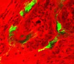
In the labeling studies, no tetracycline labeling was observed,
Furthermore, recent reports advocate a third mode of calcification,
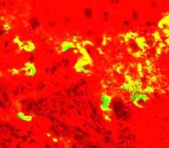
while calcein labeling was observed as a clearly defined line in
termed “transchondroid ossification”. In the present study, we
the border, but as mottled labeling with variable intensity in the
examined the modes of ossification in heterotopic bone formation
center (Figs. 1 and 2). In VG-stained section, immature matrix
and undifferentiated mesenchymal stem cells were observedscattered in the central region. Early-stage lymphocytic bone
Materials and Methods
marrow-like tissue was observed in the transitional region adjacent
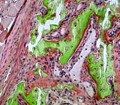
to the cartilage, calcified lamellar bone was evident in the border,
BMP was extracted by the following procedures. A block of
and many osteoids with osteoblast-like cells are present on the
bovine bone of approximately 1 mm3 was pulverized. The powder
bone marrow side. Most of the osteoblast-like cells could be
was decalcified with 0.6 N hydrochloric acid, and treated with
classified as cuboidal or intermediate type according to Villanueva.
calcium chloride and EDTA. Protein from the decalcified bone
Hematopoietic cells were observed in the bone marrow formed
was extracted using 6 M urea. The water insoluble fraction was
between the bone trabeculae, which partially became fatty marrow
collected and crude purification was conducted according to the
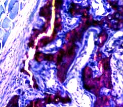
(Fig. 3). In the region adjacent to the cartilaginous matrix,
coexistence of calcified bone and osteoid was observed, and cellsmorphologically different from the ovoid osteocyte-like cells were
also found at this site (Fig. 4). At the border, the calcification
Five week-old male Std. ddy mice were used. With the animal
front is evident at the boundary between the osteoid and calcified
under Nembutal anesthesia, 5 mg of the BMP extract packed in a
bone, but the calcification front is not clear in regions with a
gelatin capsule was implanted between the fascias in the femoral
mixture of osteoid and calcified bone. In the TB-stained sections,
region. Fluorescent-labeled tetracycline (25 mg/kg) was injected
a calcification front was observed between osteoid and calcified
intraperitoneally (i.p.) after one week of implantation and calcein
bone in the border, but the calcification front was not clear in
(15 g/kg) was injected i.p. after two weeks. The animals were
areas with a mixture of osteoid and calcified bone (Figs. 5 and 6).
sacrificed at three weeks after implantation. Discussion
Heterotopic bone formation induced by BMP is generally
Non-calcified sections were prepared by the following
considered to proceed via an endochondral ossification process,
procedures. The new bone formation site was removed and
in which cartilage is formed preceding bone formation and then
immediately fixed in 70% alcohol. After fixing and staining in
replaced by bone tissue as a secondary step. Recently, however,
Villanueva bone stain (VB) solution, the tissue block was
other ossification modes have been reported. One of them is direct
embedded in methyl methacrylate. Ground sections less then 10
ossification, in which osteoblasts directly form bone matrix and
µm in thickness were prepared and stained with Villanueva
become embedded in the matrix transforming into osteocytes.
Goldner (VG) stain and toluidine blue O (TB).
Another mode has been termed the third ossification mode, andincludes “transchondroid ossification” in which “chondroid” tissue
with intermediate characteristics between bone and cartilage is
Labeling was examined with a confocal laser scanning
formed first and is subsequently replaced by bone tissue. Although
International symposium of Maxillofacial & Oral Regenerative Biology in Okayama 2005
some studies have proposed that the different ossification modes
like cells, were found adjacent to cartilaginous matrix, suggesting
are probably due to effects of the carrier, transchondroid
ossifucatuib by the transchondroid mode advocated by Yasui et
ossification has been observed irrespective of the type of carrier.
al5). These findings suggest that heterotopic bone formation
Sasano et al. used fibrous collagen membrane as carrier and
induced by BMP proceeds not only by a single ossification process,
observed cartilage with matrix expressing both type I and type II
but via at least three ossification modes. However, the BMP used
collagen. Furthermore, Kimura et al. used gelatin capsule as carrier
in the present study was a crudely purified preparation. The results
and observed immunoreactivity for type I and type II collagen in
may not be the same when synthetic human BMP is used. Further
the cytoplasm of chondrocyte-like cells and the surrounding
studies are required to examine the relation of various ossification
cartilaginous matrix on the 10th postoperative day. They confirmed
modes with cytokines such as LANKL2.
that these cells possess characteristics of both cartilage and bone,and named them chondroid bone-forming cells. The above data
References
indicate that chondroid cells possessing characteristics of both
1) Murata M., et al. Phenotype expression of BMP – induced cell
chondrocyte and osteocyte participate in ossification different from
differentiation is dependent on the matrix. Hokkaido J. Dent.
the physiological endochondral ossification.
The heterotopic bone tissue induced by our crude BMP
2) Kawakami T., et al. Transchondroid bone formation displayed
preparation showed only calcein labeling, indicating that
in BMP – induced heterotopic osteogenesis. J Hard Tissue
calcification did not occur in the first week but started at the second
week. The labeling pattern was a strongly stained line in the border
3) Sasano Y., et al. BMPs induce direct bone formation in ectopic
and mottled staining with uneven intensity in the center, suggesting
sites independent of endochondoral ossification in vivo. Anat
different modes of ossification. In the border, osteoblast-like cells
with an active morphology were observed not adjacent to
4) Kimura A., et al. Pathological examination of transchondroid
cartilaginous matrix, and TB staining demonstrated a clear
b o n e f o r m a t i o n i n d u c e d b y b o n e m o r p h o g e n e t i c
calcification front, suggesting ongoing process of direct
protein.Matsumoto Shigaku., Vol.25. No.2,3: 118-123, 1999.
ossification. In the central region where osteoid co-existed with
5) Yasui N., et al. Three modes of ossification during distraction
calcified bone, cells resembling chondroid bone-forming cells,
osteogenesis in the rat. J Bone Joint Surg., 79-B:824-830,
which were morphologically different from the ovoid osteocyte-
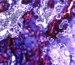
Fig. 1: Clearly demarcated line of calcein labeling
Fig. 2: Mottled calcein labeling with Fig. 3: At the border, bony matrix is
can be observed at the superficial region of the
unclear demarcation can be observed in observed, and osteoid and active
the central region of the heterotopic bone osteoblast-like cells can be seen on thetissue.
bone marrow side. O: osteoid, C: calcifiedbone
Fig. 4: In the central region, bony matrix
a n d o s t e o i d c o - e x i s t a n d c e l l s
exists, the calcification front is not sharp.
o s t e o i d a n d c a l c i f i e d b o n e .
osteocyte-like cells are also present.
: cells morphologically different from the
MEDICATION DECLARATION FORM I am an athlete and completing this form because I am: Representing Great Britain or my Home Country internationally Competing in a British Swimming, ASA, SASA or WASA National event (all disciplines, excluding masters) A new form MUST be completed annually even if the medication prescribed has not been altered or if no medication is being taken and whene
NOTas FaRmaCOTeRapéUTICas Áreas 1, 2, 3 y 7 de Atención Primaria Servicio Madrileño de Salud - CoMunidAd de MAdrid Disponible en Internet http://www.madrid.org UTIlIzaCIóN De meDICameNTOs Vol. 15 Núm. 8 Año 2008 eN el aNCIaNO CONClUsIONes: Aproximadamente el 16% de la población en • Las enfermedades crónicas, comorbilidades y España tiene 65 o más años y l


 International symposium of Maxillofacial & Oral Regenerative Biology in Okayama 2005
some studies have proposed that the different ossification modes
like cells, were found adjacent to cartilaginous matrix, suggesting
are probably due to effects of the carrier, transchondroid
ossifucatuib by the transchondroid mode advocated by Yasui et
ossification has been observed irrespective of the type of carrier.
International symposium of Maxillofacial & Oral Regenerative Biology in Okayama 2005
some studies have proposed that the different ossification modes
like cells, were found adjacent to cartilaginous matrix, suggesting
are probably due to effects of the carrier, transchondroid
ossifucatuib by the transchondroid mode advocated by Yasui et
ossification has been observed irrespective of the type of carrier.