Do you want to buy antibiotics online without prescription? https://buyantibiotics24h.net/ - This is pharmacy online for you!
Doi:10.1016/j.ajo.2004.06.040
A 43-year-old man developed bilateral painless vision
loss with photophobia, photopsia, and nyctalopia overseveral weeks. He had undergone allogeneic bone marrow
1. Strouthidis NG, Francis PJ, Stanford MR, et al. Posterior seg-
transplant 10 years earlier for aplastic anemia. He subse-
ment complications of graft versus host disease after bone marrowtransplantation. Br J Ophthalmol 2003;87:1421–1423.
quently developed GVHD with vitiligo and severe dry eyes
2. Gass JDM. Are acute zonal occult outer retinopathy and the
requiring punctal occlusion. There was no history of
white spot syndromes (AZOOR complex) specific autoim-
retinotoxic medication use and no family history of eye
mune diseases? Am J Ophthalmol 2003;135:380 –381.
disease. Four months earlier, his ophthalmologist noted
3. Gass JDM, Agarwal A, Scott IU. Acute zonal occult outer
20/20 vision with mild punctate keratopathy in each eye
retinopathy: a long-term follow-up study. Am J Ophthalmol
but no active conjunctivitis or conjunctival scarring.
Visual acuity was 20/50 in the right eye and 20/400 in
4. Jacobson SG, Morales DS, Sun XK, et al. Pattern of retinal
the left eye. The anterior segment, optic nerve, and retina
dysfunction in acute occult zonal outer retinopathy. Ophthal-
appeared normal in both eyes. Fluorescein angiography was
unremarkable. Goldmann perimetry revealed a relative
5. Jampol LM, Becker KG. White spot syndromes of the retina:
central scotoma in the right eye and a dense central
a hypothesis based on the common genetic hypothesis of
scotoma in the left eye with bilateral blind spot enlarge-
ment top). Full-field electroretinogram (ERG)
scotopic and photopic amplitudes were decreased by 20%to 30% and 50% to 75% below normal, respectively, withthe left eye more severely affected than the right. The
Two Cases of Intraoperative Anterior
first-order multifocal ERG responses from 103 hexagons ina 30-degree diameter stimulus area showed depressed
Chamber Angle Observation Using
amplitudes centrally and around the blind spot in the left
Ophthalmic Endoscope in
eye to a greater extent than the right center and
Viscocanalostomy
A complete blood count and serum vitamin A levels
Hirokazu Takahashi, MD,
were normal. No serum paraneoplastic antibodies were
Masaki Tanito, MD, PhD,
detected including those for recovering (CAR-IgG) and
Mitsunobu Yokoyama, MD,
collapsing response-mediator protein-5 (CRMP-5-IgG). Masami Park, MD, PhD, and
The patient’s central visual loss progressed but stabilized
Etsuo Chihara, MD, PhD
over 6 months, resulting in acuities of 20/200 in the righteye and 20/400 in the left eye. PURPOSE: To report endoscopic findings in the anterior
The patient’s presentation, including acute-onset
chamber angle during injection of viscoelastic material in
photopsias with bilateral asymmetric central vision loss,
viscocanalostomy (VCS).
central scotomas with blind spot enlargement, reduced
DESIGN: Observational case reports.
ERG and multifocal ERG amplitudes, and normal fun-
METHODS: Two cases of primary open-angle glaucoma
dus appearance, is consistent with Although
were treated with VCS and cataract surgery. Immediately
the exact mechanism of vision loss in patients with
before and during injection of viscoelastic material into
AZOOR remains uncertain, an autoimmune basis has
Schlemm’s canal, the anterior chamber angle was ob-
been proposed; a long-term study revealed that 28% of
served using an ophthalmic endoscope. RESULTS: In both cases, we observed viscoelastic material
corticosteroid treatment has not been effective, and
in Schlemm’s canal and leakage of viscoelastic material
autoimmune disease has not correlated with either
and blood from Schlemm’s canal into the anterior cham-
bilateral involvement or AZOOR disease No
ber away from the injection site of the viscoelastic
retinal autoantibodies have been identified in the serum
material. In one case, indocyanine green-stained vis-
Although the mechanism of vision loss in AZOOR is
coelastic material was not in Schlemm’s canal the day
unknown, a complex interplay of genetics, immune path-
after surgery.
ways, and environmental triggers has been viral infection of photoreceptors has also been The pathogenesis of both AZOOR and GVHD is incom-
Accepted for publication June 11, 2004.
From the Senshokai Eye Institute, Kyoto, Japan (H.T., M.T., M.P.,
pletely understood. Graft-versus-host disease should be
E.C.); and Kimura Eye and Internal Medicine Hospital, Hiroshima, Japan
added to the list of autoimmune conditions associated with
Inquiries to Hirokazu Takahashi, MD, Senshokai Eye Institute, Iseda-
AZOOR, and a similar mechanism may underlie the two
Cho, Minamiyama 50-1, Uji, Kyoto, 611-0043, Japan; fax: ϩ81-774-45-
FIGURE 1. Viscocanalostomy in case 1. (a) Schlemm’s canal is detected endoscopically as a red band filled with blood before the injection of viscoelastic material (arrow). (b) With viscoelastic material injection through the surgical ostia, the blood is replaced by translucent material in Schlemm’s canal, and the leakage of blood (white arrow) and viscoelastic material (arrowheads) into the anterior chamber from Schlemm’s canal is seen. (c) The ruptured site of Schlemm’s canal (large black arrow) is 3 clock-hours away from the site at which the viscoelastic material was injected (arrowhead). CONCLUSIONS: In VCS, Schlemm’s canal was filled and
bypassing juxtacanalicular connective tissue; aqueous humor was
was disrupted after viscoelastic material injection. Dis-
thought to pass through the window and the surgical ostia and
ruption of the inner wall of Schlemm’s canal and juxta-
flow into Schlemm’s Thus, accurate injection of vis-
canalicular connective tissues may contribute to the
coelastic material into Schlemm’s canal was considered critical. intraocular pressure reduction associated with VCS.
To confirm that, we observed the anterior chamber angle using
(Am J Ophthalmol 2004;138:1060 –1063. 2004 by
ophthalmic endoscope in two cases and determined the effect of
Elsevier Inc. All rights reserved.)
THEEFFICACYOFVISCOCANALOSTOMY(VCS),ANON- ● CASE 1: The right eye of a 71-year-old woman with
perforating surgery introduced by Stegmann and
primary open-angle glaucoma was treated with cataract
assoin reducing intraocular pressure (IOP) in glaucoma-
surgery and VCS. Just before viscoelastic material injec-
tous eyes was reported with VCS and combined with
tion, a fiberoptic endoscope (FV-2000, Tokyo Denshi,
cataract Viscocanalostomy includes deroofing of
Tokyo, Japan) was inserted into the anterior chamber
Schlemm’s canal, preparation of a “window” formed by Descem-
through the corneal side port and Schlemm’s canal was
et’s membrane, creation of a scleral “lake,” and injection of
visualized When the viscoelastic material was
viscoelastic material into the cut ends of Schlemm’s canal
injected into the surgical ostia, Schlemm’s canal ruptured,
“surgical ostia” to maintain the lumen of Schlemm’s canal and
and we observed viscoelastic material and blood leaking
prevent healing of the surgical Stegmann and associates
from the rupture site apart from the surgical
postulated that VCS facilitates aqueous humor outflow by
FIGURE 2. Viscocanalostomy in case 2. (a) When viscoelastic material was stained with indocyanine green, displacement of the blood in Schlemm’s canal by the green material is seen more clearly by endoscopy of the anterior chamber angle (arrow). (b) With the injection of additional viscoelastic material, Schlemm’s canal (seen as a green band) is distended (arrowheads), and the leakage of the viscoelastic material into the anterior chamber is observed clearly (arrow). (c) The site of leakage of the viscoelastic material from Schlemm’s canal (arrow) is 3 clock-hours away from the injection site of the viscoelastic material (arrowhead).
● CASE 2: The left eye of an 80-year-old woman with
green might have diffused out, suggesting that viscoelas-
primary open-angle glaucoma was treated with cataract sur-
tic material does not remain long enough to prevent
gery and VCS. To improve visibility, we used viscoelastic
healing of the surgical ostia. Additionally, other inves-
material stained with indocyanine green at a concentration of
tigators reported the insufficient permeability of De-
1 mg/ml. When viscoelastic material was injected, Schlemm’s
scemet’s membrane against water to relieve elevated
canal filled and when additional material was
IOP in glaucomatous Thus, other mechanisms of
injected, it ruptured. We clearly observed the leakage of
IOP reduction may occur during VCS other than those
viscoelastic material into the anterior chamber
reported by Stegmann and associates.
from a site apart from the surgical ostia With a
We observed the leakage of viscoelastic material and blood
gonioscopic examination, green material was not observed in
reflux from Schlemm’s canal, suggesting the rupture of inner
Schlemm’s canal 1 day postoperatively.
wall of Schlemm’s canal. Viscocanalostomy may act as a
Our observations confirm that more than a quarter of
trabeculotomy, allowing aqueous humor to pass into
Schlemm’s canal from the surgical ostia was filled with
Schlemm’s canal through its ruptured inner wall and juxta-
viscoelastic material. Schlemm’s canal in monkey and
canalicular connective tissue, as we and others
human autopsy eyes was dilated with viscoelastic
In conclusion, Schlemm’s canal was distended by
In case 2, the dilation was confirmed in a living human eye
viscoelastic material during VCS. Disruption of the
inner wall of Schlemm’s canal and juxtacanalicular
In case 2, the green material in Schlemm’s canal was
connective tissue may contribute to IOP reduction
not detected one day postoperatively, thus indocyanine
CAT-SCRATCHDISEASE(CSD)ISASELF-LIMITEDINFEC-
tion caused by Bartonella hensalea. We describe a CSD
1. Stegmann R, Pienaar A, Miller D. Viscocanalostomy for
patient who presented with unilateral panuveitis and
open-angle glaucoma in black African patients. J Cataract
papillitis, diffuse choroidal thickening on ultrasound, and
angiographic findings suggestive of Vogt-Koyanagi-Harada
2. Sunaric-Megevand G, Leuenberger PM. Results of viscocana-
lostomy for primary open-angle glaucoma. Am J Ophthalmol
(VKH) disease. This case highlights another posterior
3. Tanito M, Park M, Nishikawa M, Ohira A, Chihara E.
A 54-year-old Mestizo woman presented with acute,
Comparison of surgical outcomes of combined viscocana-
painless loss of vision in her right eye of 5 days’ duration
lostomy and cataract surgery with combined trabeculotomy
and admitted to headache and tinnitus. Best-corrected
and cataract surgery. Am J Ophthalmol 2002;134:513–520.
visual acuity was counting fingers at 6 feet in the right
4. Johnson DH, Johnson M. Glaucoma surgery and aqueous
eye and 20/25 in the left eye. There was no relative
outflow: how does nonpenetrating glaucoma surgery work?
afferent pupillary defect. Slit-lamp examination showed
ϩ1 anterior chamber cells and ϩ2 vitreous cells in the
5. Fatt I. Permeability of Descemet’s membrane to water. Exp
right eye. Dilated fundoscopic examination of the right
eye revealed optic disk edema with a hemorrhageinferior temporally, choroidoretinal folds, and peripap-illary edema The left eye was normal. Bartonella henselae Infection
Fluorescein angiography showed leakage around the
Presenting as a Unilateral Panuveitis
optic nerve and multiple hyperfluorescent spots with adja-cent leakage A linear radial hyperfluorescent
Simulating Vogt-Koyanagi-Harada
streak and adjacent hypofluorescence confirmed the pres-
Syndrome
ence of choroidal folds. Echographic examination revealed
Rahul N. Khurana, MD, Thomas Albini, MD,
diffuse medium reflective thickening of the choroid of theright eye only. Ronald L. Green, MD, Narsing A. Rao, MD, and
Initial laboratory tests including a complete blood cell
Jennifer I. Lim, MD
count, chemistry profile, reactive plasma reagin, angioten-sin-converting enzyme, and lyme titers were normal. Com-
PURPOSE: To report an unusual ocular manifestation of
puted tomography scan of the brain was unremarkable. cat scratch disease.
Based on angiographic and ultrasound findings, we enter-
DESIGN: Observational case report.
tained the diagnosis of VKH. However, cerebrospinal fluid
METHODS: Review of the clinical, laboratory, photo-
(CSF) studies revealed normal cell counts with no leuko-
graphic, and angiographic records of a patient with cat
cytosis or neoplastic cells. Purified protein derivative
scratch disease.
(PPD) was positive, but chest X-ray was negative. RESULTS: A 54-year-old woman presented with counting
Three weeks later, visual acuity improved to 20/80. fingers visual acuity in the right eye associated with optic
Immunoglobulin (Ig) G serum titers for B. hensalae were
disk edema, diffuse choroidal thickening, and panuveitis.
obtained and elevated at 1:256; IgM titers were nega-
Fluorescein angiography showed disk leakage and hyper-
tive. She was diagnosed with CSD and treated with a
fluorescent spots with late leakage suggestive of Vogt-
2-week course of doxycycline. She recalled exposure to
Koyanagi-Harada disease. She was diagnosed with cat
kittens but denied being scratched. Seven weeks after
scratch disease by serum antibody titers and clinical
presentation, visual acuity was 20/25, the papillitis and
choroidal folds resolved, and echography showed no
CONCLUSIONS: Ocular manifestations of cat scratch disease can include diffuse thickening of the choroid.
Vogt-Koyanagi-Harada (VKH) syndrome consists of
Cat scratch disease may manifest with angiographic
panuveitis associated with neurologic or cutaneous man-
features suggestive of Vogt-Koyanagi-Harada disease. (Am J Ophthalmol 2004;138:1063–1065. 2004 by
mon. In unilateral cases, the second eye is affected
Elsevier Inc. All rights reserved.)
within 2 weeks. The patient initially presented withunilateral panuveitis, optic disk edema, choroidal thick-ening, and fluorescein angiographic findings consistent
Accepted for publication June 11, 2004.
with VKH. Even though VKH is a bilateral disease,
From the Doheny Eye Institute, University of Southern California
Keck School of Medicine, Los Angeles, California.
unilateral occur, and the patient’s pre-
Supported in part by NIH EY 03040 and an unrestricted grant from
sentation raised this possibility. Her negative workup for
VKH and her spontaneous improvement are consistent
Inquires to Jennifer I. Lim, MD, Los Angeles, CA 90027; fax: (323)
Comunicado de Prensa Descubre cómo lucir una cabellera saludable y estilizada con Smooth ‘N Shine • La marca Smooth ’N Shine presenta su nuevo mousse Diamond Luster Anti-Frizz , así como la nueva imagen de su línea actual de mousses. • Su innovadora acción termo-protectora ayuda a sellar la estructura capilar, protegiendo el cabello contra el calor.
Fertility Physicians of Northern California Fertility Physicians of Northern California is participating in a multicenter ovulation induction research study, sponsored by Serono, Inc. This study will evaluate the safety and tolerability of anastrozole in women who do not ovulate regularly, by comparing it to clomiphene citrate, often known as Clomid® or Serophene®. It also aims to determine an
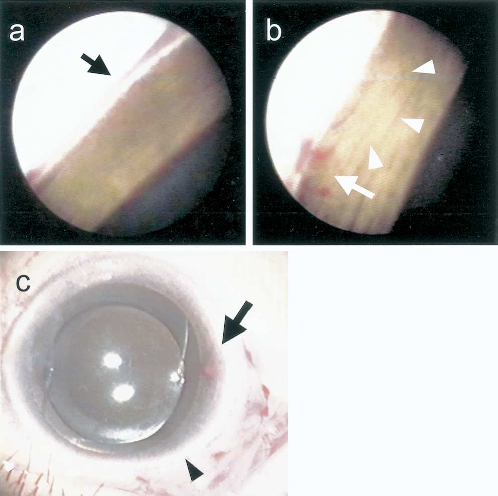 FIGURE 1. Viscocanalostomy in case 1. (a) Schlemm’s canal is detected endoscopically as a red band filled with blood before the
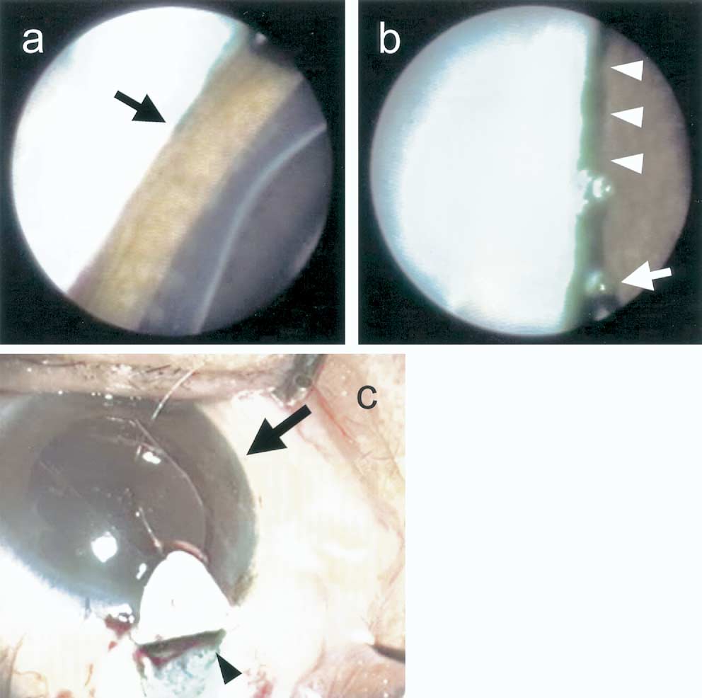 FIGURE 2. Viscocanalostomy in case 2. (a) When viscoelastic material was stained with indocyanine green, displacement of the