Do you want to buy antibiotics online without prescription? https://buyantibiotics24h.net/ - This is pharmacy online for you!
Jalilian.indd
ORIGINAL PAPER Development of a radiolabeled Amir R. Jalilian, Mahdokht Jouiaei, glucagon compound for imaging Ali R. Doroudi, Fatemeh Bolourinovin, Javad Garousi Abstract. In order to develop a possible Ga-labeled glucagon (GCG) compound for imaging studies, biosynthetic glucagon (GCG) was labeled with [67Ga]-gallium chloride after conjugation with freshly prepared diethylenetriamine- pentaacetic acid dianhydride (ccDTPA). After solid phase purification of the radiolabeled hormone, high performance liquid chromatography (HPLC) and instant thin-layer chromatography (ITLC) showed a radiochemical purity around 95% in optimized conditions (specific activity = 296–370 GBq/mM; labeling efficiency 85%). Preliminary in vivo studies (ID·g–1%) in male wild-type rats showed heart : muscle, liver : muscle, lung : muscle and stomach : muscle ratios to be 5.53, 2.9, 7.56, 3.69, 3.2 (in 5 min), respectively while after 2 h liver : blood, lung : blood and spleen : blood ratios were 14.21, 16.86 and 7.8, respectively. The data suggests 5 min post injection, the tracer is accumulated in GCGR rich tissues which is in agreement with biodistribution studies and reported GCG receptors (GCGRs). The results of the present study can possibly offer a candidate for non-invasive imaging of glucagon receptor related diseased and malignancies such as glucagonoma. Key words: glucagons • radiolabeling • biodistribution • Ga-67 Introduction
Glucagon (GCG) is a linear peptide of 29 amino acids. Its primary sequence is almost perfectly conserved among vertebrates. GCG helps to maintain the level of glucose in the blood by binding to glucagon receptors (GCGRs) on hepatocytes, causing the liver to release glucose, stored in the form of glycogen, through a pro-cess known as glucogenolysis.
125I-GCG is the only radiolabeled GCG compound
that has been reported in the literature according to our knowledge and is frequently used in radio-pharma-cological studies. 125I-GCG has been used in the study of GCG hydrolysis by proximal tubules, identification
A. R. Jalilian, F. Bolourinovin, J. Garousi
of renal extraction mechanisms [20], GCG receptor
Radiopharmaceutical Research and Development
binding [7], rat brain binding [9], reabsorption measure-
ments in urinary tract [4] and hormone internalization
Nuclear Science and Technology Research Institute
in hepatocyte [3]. Also the photoreactive 125I-GCG was
The presence of GCGRs in various human ma-
Tel.: +98 21 8822 1103, Fax: +98 21 8822 1105,
lignancies has been well documented. For instance,
glucagonoma is a neuroendocrine tumour that develops
from glucagon-producing pancreatic cells. They are usu-
ally slow-growing, but generally advanced at diagnosis,
Ahvaz Jundishapur University of Medical Sciences,
and metastatic disease is virtually incurable. Liver is the
Golestan blvd., Postal code: 61357-33184, Ahvaz, Iran
most common site of metastatic disease [19].
The diagnosis of pancreatic endocrine tumors, such
as glucagonomas, is difficult and requires a careful his-
tory and examination combined with laboratory tests
and radiologic imaging. Surgical resection remains
the treatment of choice even in the face of metastatic disease. Further development of novel diagnostic
and treatment modalities offers potential to greatly improve quality of life and prolong disease-free sur-
68Zn(p,2n)67Ga was used as the best nuclear reaction for
vival for patients with pancreatic endocrine tumors [5].
the production of 67Ga. Impurities could be removed
Due to overexpression of GCGRs on these malignant
in a radiochemical separation process. After the target
cells, the development of an appropriate radiolabeled
bombardment process, chemical separation was carried
compound capable of nuclear medicine imaging using
out in no-carrier-added form. The irradiated target was
single photon emission computed tomography (SPECT)
dissolved in 10 mol·L–1 HCl (15 ml) and the solution was
and positron emission tomography (PET) can be of
passed through a cation exchange resin (AG 50W, H+
great importance. According to our knowledge, there
form, mesh 200–400, h: 10 cm, Ø: 1.3 cm) which had
were no reports of radiolabeled GCG for imaging studies
been preconditioned by passing 25 mL of 9 mol·L–1 HCl.
in the literature. In this work, following the preparation
The column was then washed by 25 mL of 9 mol·L–1 HCl
of a GCG conjugate for the use in diagnostic GCGR
at a rate of 1 mL/min to remove copper and zinc ions.
studies. 67Ga-GCG was prepared and used for pre-
To the eluent 30 mL of water plus about 100 mL of a
liminary biodistribution studies, based on our recent
6 mol·L–1 HCl solution was added. The latter solution
experiences on the preparation of radiometal-labeled
was loaded on another exchange resin (AG1X8 Cl–
form, 100–200 mesh, h: 25 cm, Ø: 1.7 cm) pretreated with 6 mol·L–1 HCl (100 mL). Finally, the gallium-67 was eluted as [67Ga]GaCl3 using 2 mol·L–1 HCl (50 mL);
Experimental
Production of 67Ga was performed at the Agricultural,
Gamma spectroscopy of the final sample was carried
Medical and Industrial Research School (AMIRS,
out by counting the activity in a high-purity germanium
Karaj, Iran) using a 30 MeV cyclotron (Cyclone-30,
(HPGe) detector coupled to a Canberra™ multichannel
IBA, Belgium). Enriched zinc-68 chloride (enrichment
> 95%) was obtained from the Ion Beam Separation Department at AMIRS. All chemicals were purchased
from commercial sources. GlucaGen® (glucagon [rDNA origin] for injection) manufactured by Novo Nordisk
The presence of zinc and copper cations were checked
A/S (1 mg/ml, 1 IU/ml) and was used without further
by the polarography method. The area under curve of
purification. Cyclic DTPA dianhydride was freshly pre-
polarogram of the test samples were lower than the stan-
dards even at 1 ppm of standard zinc and copper [15].
chromatography (ITLC) was performed by counting Whatman no. 2 papers using a thin-layer chromatog-
Conjugation of ccDTPA with human recombinant hCG
raphy scanner, Bioscan AR2000, Bioscan Europe Ltd. (France). Analytical HPLC to determine the specific
The chelator ccDTPA was conjugated to the GCG using
activity was performed by a Shimadzu LC-10AT (Japan)
a small modification of the well-known cyclic anhydride
instrument, armed with two detector systems, a flow
method [8]. Conjugation was performed at a 1:1 molar
scintillation analyzer (Packard-150 TR, USA) and a UV-
ratio. In brief, 20 μl of a 1 mg·ml–1 suspension of DTPA
-visible (Shimadzu, Japan) using Whatman Partisphere
anhydride in dry chloroform (Merck, Germany) was
C-18 column (250 × 4.6 mm), Whatman, USA. Solid
pipetted under ultrasonication and transferred to a glass
phase purification of the radiolabeled hormone was
tube. The chloroform was evaporated under a gentle
stream of nitrogen. Commercially available GCG (1 mg,
Calculations were based on the 184 keV peak for 67Ga.
1 mL, pH 6, ≈ 0.3 nmol–1) was subsequently added and
All values were expressed as mean ± standard devia-
gently mixed at room temperature for 60 min followed
tion and the data were compared using Student’s t-test.
Animal studies were performed in accordance with the United Kingdom Biological Council’s Guidelines on
Radiolabeling of GCG conjugate with 67Ga
the Use of Living Animals in Scientific Investigations, 1987. The approval of AMIRS Ethical Committee was
The GCG conjugate was labeled using an optimized
obtained for conducting this research. The wild-type
protocol according to the literature [14].Typically,
rats (NMRI) were purchased from the Pasteur Insti-
74 MBq of 67Ga-chloride (in 0.2 mol·L–1 HCl) was added
tute of Iran, Karaj, all weighing 180–200 g; they were
to a conical vial and dried under a flow of nitrogen.
acclimatized at a proper rodent diet and 12 h/12 h day/
To the 67Ga containing vial, the conjugated fraction
night light/darkness. The percentage of injected dose in
was added in 1 mL of phosphate buffer (0.1 mol·L–1,
tissue (ID·g–1%) were determined using a high-purity
pH 6) and mixed gently for 30 s. The resulting solution
germanium (HPGe) detector coupled with a Canberra™
was incubated at room temperature for 30 min. Fol-
(model GC1020-7500SL, USA) multichannel analyzer
lowing incubation, the radiolabeled GCG conjugate
based on the area under the curve for 184 keV photo-
was checked using for purity the ITLC/RTLC methods.
peak and calculated efficiency of the counting system.
In the case of presence of unreacted amounts of im-
Development of a radiolabeled glucagon compound for imaging
purities, the sample can be purified using solid phase extraction using C18 Sep-Pak. Briefly, the column was pretreated with absolute ethanol (3 mL) and water (2 mL), respectively followed by the injection of radiolabeling mixture. The column was left at room temperature for 5 min and then was washed with water
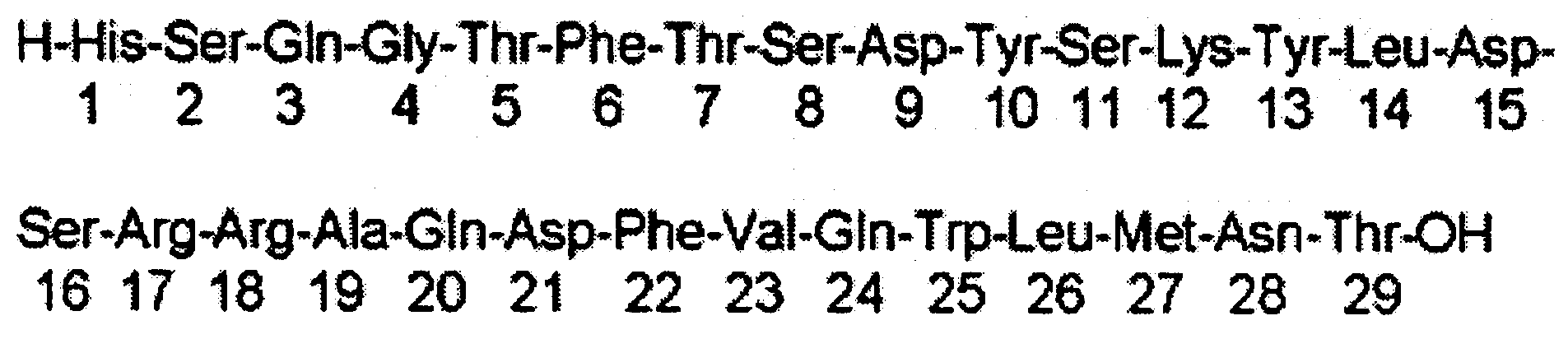
Fig. 1. Amino acid sequence of GCG.
fractions (1 mL) till the flow-through activity in each fraction was less than 10 μCi. Finally, the radiolabeled compound was eluted from the column using 1 mL
fractions of citrate buffer (pH 5.5). Control labeling experiments were also performed using 67GaCl
Glucagon with a molecular weight of 3483, is a single-
-chain polypeptide containing 29 amino acid residues
(isoelectric point pI 7) is synthesized and secreted from
A cells of pancreatic islets scattered throughout the islet. The liver and kidney seem to be the major sites
Paper chromatography. A 5-μL sample of the final frac-
of glucagon catabolism, but the relative contribution of
tion was spotted on a chromatography paper (Whatman
no. 2, Whatman, UK), and developed in a mixture of
In this work, the labeling yield of 67Ga-DTPA-GCG
has been studied in a wide range of GCG/DTPA ratios
High performance liquid chromatography. HPLC
in order to optimize the process and to improve 67Ga-
was performed on the final preparation using acetate
-DTPA-GCG performance in vitro. The overall ra-
buffer solution (50 Mmol·L–1 pH 5.5) as eluent A
diolabeling efficiency was over 85%. Because of its
(flow rate: 1 ml/min) for 20 min in order to elute low
isoelectric point (IEP) of around 7, GCG is soluble in
molecular mass components. Radiolabeled peptide
lower physiological serum pH (5.5–6) being adequately
was eluted using a gradient of the latter solution
stable hypothetically [18]. Figure 1 demonstrates the
(100 to 0%) and citrate buffer solution B (50 mM,
peptide sequence for GCG and considering the exis-
pH 4,0 to 100%, 5 min A;100%, B;0%, 5 min A;70%, B;30,
tence of one lysine moiety in the structure, the NH2
5 min A;50%, B;50%, 50 A:0%, B;100%) using reverse
mediated conjugation through ccDTPA acylation
looked feasible, leading to a possible 1:1:1 stoichiom-etry of the DTPA:GCG:Ga ratio, which was a suitable
Stability testing of the radiolabeled compound
The protein was conjugated using ccDTPA in a
Stability of 67Ga-DTPA-GCG in phosphate buffer
similar way already reported, followed by size exclusion
solution was determined by storing the final solution
chromatography of the compound showing 78–85%
at 4°C for 4 h and performing frequent ITLC analy-
radiochemical purity after 1 h. Due to the relative insta-
sis to determine radiochemical purity. ITLC analysis
bility of the radiolabeled peptide at room temperature
of the conjugated product was also performed to moni-
instead of increasing time to obtain higher purities,
tor degradation products or other impurities after the
solid-phase extraction using C18 column was used. The
conjugated DTPA-GCG was stored at –20°C for more
radiolabeled mixture was loaded on the preconditioned
than 1 month. After subsequent 67Ga-labeling of the
C18 Sep-Pak. Eluting the loaded column with water,
stored conjugated product, both labeling efficiency and
removed free 67Ga3+ as well as 67GaDTPA due to their
ionic properties. After purging the column with nitrogen for 5 min, the radiolabeled protein was eluted using
Stability testing of the radiolabeled compound in presence
citrate buffer in the first 3 elutions (1 mL).
The eluted fractions were checked for the pres-
ence of radioactivity in order to determine the 67Ga-
Labeled compound stability in serum, was assessed by
-DTPA-GCG containing fractions. The fraction with a
gel filtration on a Sepharose column (1 × 30 cm). The
maximum radioactivity was chosen as the suitable final
column was equilibrated with PBS and eluted at a flow
product for quality control and with appropriate specific
rate of 0.5 mL·min–1 at room temperature; 0.5 mL frac-
At this stage, the buffer eluted fraction with the high-
est activity was tested by ITLC and HPLC in order to
Biodistribution of 67Ga-DTPA-GCG in wild-type rats
determine the radiochemical purity before administra-tion to wild-type rats for biodistribution studies. Figure 2
To determine its biodistribution, 67Ga-DTPA-GCG was
shows the ITLC chromatograms for free 67Ga3+ and the
administered to wild-type rats. A volume (50 μl) of
labeled compound after solid phase extraction.
final 67Ga-DTPA-GCG solution containing 40±2 μCi
Figure 3 demonstrated the HPLC chromatogram of
radioactivity was injected intravenously to rats through
67Ga3+ which was tested as a control. In HPLC experi-
ments of the radiolabeled compound, two major peaks
The animals were sacrificed at exact time intervals
can be observed. The fast eluting component (2.79 min)
(5, 15 min, 1, 2, 4, 24, 48 and 72 h). The specific activ-
was shown to be a mixture of free 67Ga and 67GaDTPA
ity of different organs was calculated as percentage of
which was washed out on the reverse phase stationary
urea under the curve of 184 keV peak per gram using
phase. The radiolabeled protein was washed out at
Fig. 2. ITLC of free 67Ga used in the radiolabeling (right) and 67Ga-GCG solution (left) on Whatman paper using 10 mM DTPA solution as eluent.
Considering the amount of activity used (74 MBq)
showed that 25% of the radioactivity is not eluted
and the radiochemical purity of the final purified sample
in the same fraction. Thus, there is a fraction of the
(95%), a specific activity of 22–23 TBq·mmol–1 has been
tracer which has degraded or transchelated 67Ga to
other serum proteins over this time period. Also the
The stability of the radiolabeled protein in vitro was
biodistribution data supports this observation.
determined after challenge with phosphate-buffered
The distribution of free 67GaCl3 in appropriate buf-
saline and serum. ITLC analysis showed that the pro-
fer has been already reported elsewhere [12]. Figure 5
teins retained the radiolabel over a period of 1 h in the
demonstrates the biodistribution of [67Ga]-DTPA-GCG
These results were confirmed by gel filtration chro-
A volume (0.1 ml) of final [67Ga]-DTPA-GCG solu-
matography. After incubation of [67Ga]-DTPA-GCG
tion containing 40 μCi of radioactivity was injected into
with PBS for 2 h, there was no change in the Rf for
the rats’ dorsal tail vein. The total amount of radioactiv-
[67Ga]-DTPA-GCG and also there was no evidence for
ity injected into each rat was measured by counting the
a large-scale release of free Ga resulting in the appear-
1 mL syringe before and after injection in a dose calibra-
Gel filtration chromatography of [67Ga]-DTPA-
The animals were sacrificed by CO2 asphyxiation at
-GCG after incubation for 2 h with human serum
selected times after injection (5 min – 72 h), the tissues (blood, heart, spleen, kidneys, liver, intestine, muscle, bone, brain, stomach, lung, skin, fat, pancreas and bladder) were weighed and their specific activities were
Fig. 3. HPLC chromatogram of free 67GaCl Fig. 4. HPLC chromatogram of SPE purified final radiola-
reversed phase column using a gradient of acetate/citrate
beled solution on a reversed phase column using a gradient
Development of a radiolabeled glucagon compound for imaging
Fig. 5. Biodistribution of [67Ga]-DTPA-GCG (1.85 MBq, 40 μCi) in wild-type rats 5 min–72 h after IV injection via tail vein (ID·g–1%: percentage of injected dose per gram of tissue calculated based on the area under curve of 184 keV peak in gamma spectrum).
determined with a γ-ray scintillation detector as a
of glucagon. Parenteral administration of glucagon pro-
percent of area under the curve of 184 keV per gram
duces relaxation of the smooth muscle of the stomach,
duodenum, small bowel and colon. This indirectly pro-
The tracer is removed from the blood stream after
poses the existence of GCGRs in GI tract, as it can be
1 h and this is in accordance with biodistribution pattern
obviously observed in stomach 48–72 h post injection.
for most radiolabeled small proteins and peptides.
Five min post injection, the heart:muscle, liver:muscle,
Glucagon receptors are mainly expressed in liver
lung:muscle and stomach: muscle ratios were 5.53, 2.9,
and in kidney with lesser amounts found in heart, adi-
7.56 and 3.69, respectively while after 2 h the liver:blood,
pose tissue, spleen, thymus, adrenal glands, pancreas,
lung:blood and spleen: blood ratios were 14.21, 16.86
cerebral cortex, and gastrointestinal tract.
Heart uptake demonstrates a significant uptake
The half-life of glucagon in plasma is approximately 3
after 5 min post injection (3–4%), but due to possible
to 6 min [10], while under the circumstances in IV injec-
degradation as reported [2], the accumulation decreases
tions it has been reported to be 25–30 min [11], thus the
main receptor binding takes place in the first 1–20 min
Liver is a high uptake organ possibly due to two
post injection, although the accumulation at longer time
different mechanisms; a) the presence of high GCGRs
intervals at the receptor rich tissues is also observed. This
in the hepatocytes mediating the glycogenolysis and
can be possibly caused by the unknown cell accumulation
b) liver is repeatedly reported as the major uptake
of the tracer and/or the metabolites. Also a slight rat
tissue for proteins and some other macromolecules,
brain uptake can be observed on 15 min post injection
this bi-mechanistic pattern can also be supported by a
(1–2%) which is in accordance with previous reports
significant decrease in liver uptake after 1 h. 5–15 min
[9]. Although glucomoma has been known for some
post injection, the uptake is increasing and this can be
time past, the diagnosis has not been well established
due to the direct receptor : ligand interaction, but after
yet. Arterial stimulation and venous sampling (ASVS) is
1 h the 2 d increase is possibly caused by non-specific
known to be useful for insulinoma and gastrinoma, and
protein uptake in the liver. Degradation processing may
just recently its usefulness for glucagonoma has been
occur locally in target tissues such as the pancreas, liver
verified using this invasive method followed by sampling
or heart, as well as in the circulation [14].
and tissue studies [17]. The results of the present study
It has long been known that the kidney is capable of
can possibly offer a candidate for non-invasive imaging
degrading glucagon. Arteriovenous gradients across the
of glucagon receptor related diseased and malignancies
kidney in normal animals infused with glucagon indicate
such as glucagonoma. Also the early diagnosis of this
extraction of 23 to 39% of the presented glucagon [1, 19].
malignancy is a major breakthrough for the therapy,
Because less than 2% of the extracted hormone appears
while it has just recently been reported that this malig-
in urine and because nonfiltering kidneys continue to
nancy can develop hepatic metastasis [16]. The results
extract appreciable amounts of glucagon, it seems that
of the present study can possibly offer a candidate for
both tubular re-absorption and postglomerular capillary
non-invasive imaging of glucagon receptor related
tubular uptake precede renal parenchymal degradation
diseases and malignancies such as glucagonoma.
Although many radioiodine labeled glucagons are
References
reported for in vitro studies, none of them contain I-123 compound, suitable for imaging while this compound
1. Bastl C, Finkelstein FO, Sherwin R, Hendler R, Felig P,
can be a good candidate as well. Considering the
Hayslett JP (1977) Renal extraction of glucagon in rats
biological half-life of native glucagon (25–30 min) and
with normal and reduced renal function. Am J Physiol
intermediate I-123 half-life (13.2 h), the radiolabeling of
the GCG with radioiodine with I-123 does not seem ap-
2. Blache P, Kervran A, Le-Nguyen D et al. (1993) Endopep-
tidase from rat liver membranes, which generates miniglu-
propriate, while Ga-68, a widely used PET radionuclide
cagon from glucagon. Biol Chem 268;29:21748–21753
(half-life 68 min), seems an interesting candidate for
3. Canivet B, Gorden P, Carpentier JL, Orci L, Freychet P
developing a tracer. Thus, in this work the radiolabel-
(1981) Glucagon degradation in isolated rat hepatocytes:
ing GCG performed using Ga-67 radionuclide due to
effect of ammonium chloride and chloroquine. Mol Cell
availability in our center. The optimized method and
conditions can easily be used for Ga-68 labeling.
4. Carone FA, Peterson DR, Flouret G (1982) Renal tubular
processing of small peptide hormones. J Lab Clin Med 100;1:1–14
Discussion
5. Davies K, Conlon KC (2009) Neuroendocrine tumors of the
pancreas. Review. Curr Gastroenterol Rep 11;2:119–127
6. Demoliou-Mason C, Epand RM (1982) Identification of
Total labeling and formulation of [67Ga]-DTPA-GCG
the glucagon receptor by covalent labeling with a radio-
took about 60 min. A suitable specific activity product
labeled photoreactive glucagon analogue. Biochemistry
was formed via insertion of 67Ga cation. No other
labeled conjugates were observed upon ITLC and/or
7. Hagopian WA, Tager HS (1984) Receptor binding and cell-
HPLC analysis of the final preparations. The radiola-
-mediated metabolism of [125I]monoiodo-glucagon by iso-
beled complex was stable in human serum for at least
lated canine hepatocytes. J Biol Chem 259;14:8986–8993
1 h and no significant amount of free 67Ga as well as
8. Hnatowich DJ, Layne WW, Child RL (1983) Radioac-
67Ga-DTPA was observed. A radiochemical purity of
tive labeling of antibody: a simple and efficient method.
95% was detected by HPLC. The final preparation
was administered to wild-type rats and biodistribution
9. Hoosein NM, Gurd RS (1984) Identification of glu-
cagon receptors in rat brain. Proc Natl Acad Sci USA
of the radiopharmaceutical was checked 5 min to 72 h
later. Preliminary in vivo studies (ID·g–1%) in male
10. http://www.rxmed.com/b.main/b2.pharmaceutical/
wild-type rats showed a significant heart and liver
uptake of the tracer after 5 min, in agreement with the
11. Information for the physician glucagon for injection (rDNA
biodistribution studies and reported GCG receptors
origin) PA 2284 AMP, Description. Literature revised
(GCGRs). Tissue : muscle values extracted from tissue
February 18, 2005, Eli Lilly and Company, Indianapolis,
accumulated activities demonstrate that 5 min post
injection the tracer is possibly accumulated in GCGR
12. Jalilian AR, Mehdipour P, Akhlaghi M, Yousefnia H,
rich tissues. [67Ga]-DTPA-GCG can be a suitable probe
Shafaii K (2009) Evaluation of a [67Ga]-thiosemicarbazone
for biodistribution study of CGR in various physiologi-
complex as tumor imaging agent. Sci Pharm 77:343–354
13. Jalilian AR, Mirsadeghi L, Haji-Hosseini R (2007) Prepa-
cal and malignant diseases with over-expressed CGRs.
ration and biodistribution of [67Ga]-DTPA-rituximab in
Due to interesting characteristics of 68Ga radionuclide
normal rats. J Radioanal Nucl Chem 274:175–179
(half-life 68 min) in molecular imaging, and biological
14. Jalilian AR, Rowshanfarzad P, Shafaii K et al. (2005)
half-life of GCG, developing a 68Ga-labeled tracer can
Development of 111In-DTPA-human polyclonal antibody
be of great interest. The results of the present study can
complex for long-term inflammation/infection detection.
possibly offer a candidate for non-invasive imaging of
glucagon receptor related diseased and malignancies
15. Marczenko Z (1976) Spectrophotometric determination of
elements. John Wiley & Sons Inc, New York, pp 238–240
16. Obi N, Katabami T, Obi R, Odanaka M, Sasano K, Tanaka
Y (2009) Primary malignant hepatic glucagonoma: an autopsy case. Endocr J 56;5:715–719
Acknowledgment. The authors wish to thank Mr A. A.
17. Okauchi Y, Nammo T, Iwahashi H et al. (2009) Gluca-
Rajamand for 67Ga production and Mr S. Daneshvari for
gonoma diagnosed by arterial stimulation and venous
conducting animal studies. We would also like to thank
sampling (ASVS). Intern Med 48;12:1025–1030
AEOI grant (4/2/2/1/6, 2005) for supporting this project.
18. Peterson DR, Green EA, Oparil S, Hjelle JT (1986)
Transport and hydrolysis of glucagon in the proximal nephron. Am J Physiol 251(3 Pt 2):F460–F467
19. Pinto Marín A, Hernández Agudo E, Feliú J, González
Barón M (2009) Pancreatic glucagonoma presenting as a pulmonary mass. Clin Transl Oncol 11;1:60–62
20. Righetti PG, Tudor G, Ek K (1981) Isoelectric points and
molecular weights of proteins. J Chromatogr 220:115–194
Professor and Kentucky Center for School Safety Fellow Department of Safety, Security, and Emergency Management Eastern Kentucky University 521 Lancaster Avenue Richmond, KY 40475 Office- 859-622-6681 Fax- 859-622-6650 email- david.may@eku.edu Home Page- http://www.corrections.eku.edu/May/David_May.htm EDUCATION Ph.D. Sociology, Mississippi State University, Mississippi State, MS, December 1997
CONFIDENTIAL TRAVEL VACCINATION QUESTIONNAIRE For travel ing abroad you may require vaccinations and health advice. Please complete this questionnaire and hand it in at reception . Please telephone after 48 hours to ascertain if a travel consultation is necessary . It is advisable to complete this form 6 – 8 weeks prior to travel ing as multiple appointments may be required. Please c
 Development of a radiolabeled glucagon compound for imaging
purities, the sample can be purified using solid phase extraction using C18 Sep-Pak. Briefly, the column was pretreated with absolute ethanol (3 mL) and water (2 mL), respectively followed by the injection of radiolabeling mixture. The column was left at room temperature for 5 min and then was washed with water
Fig. 1. Amino acid sequence of GCG.
Development of a radiolabeled glucagon compound for imaging
purities, the sample can be purified using solid phase extraction using C18 Sep-Pak. Briefly, the column was pretreated with absolute ethanol (3 mL) and water (2 mL), respectively followed by the injection of radiolabeling mixture. The column was left at room temperature for 5 min and then was washed with water
Fig. 1. Amino acid sequence of GCG.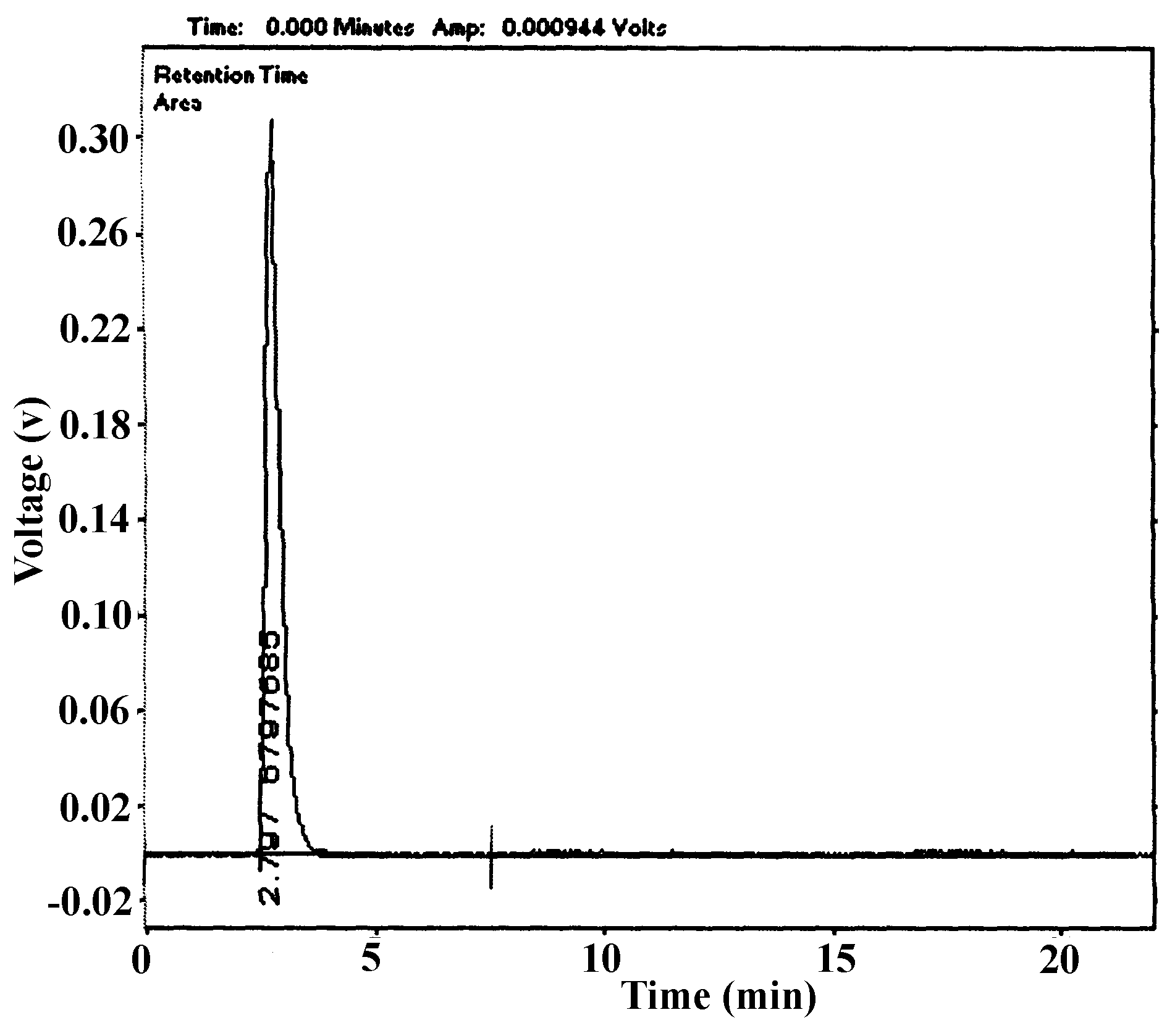
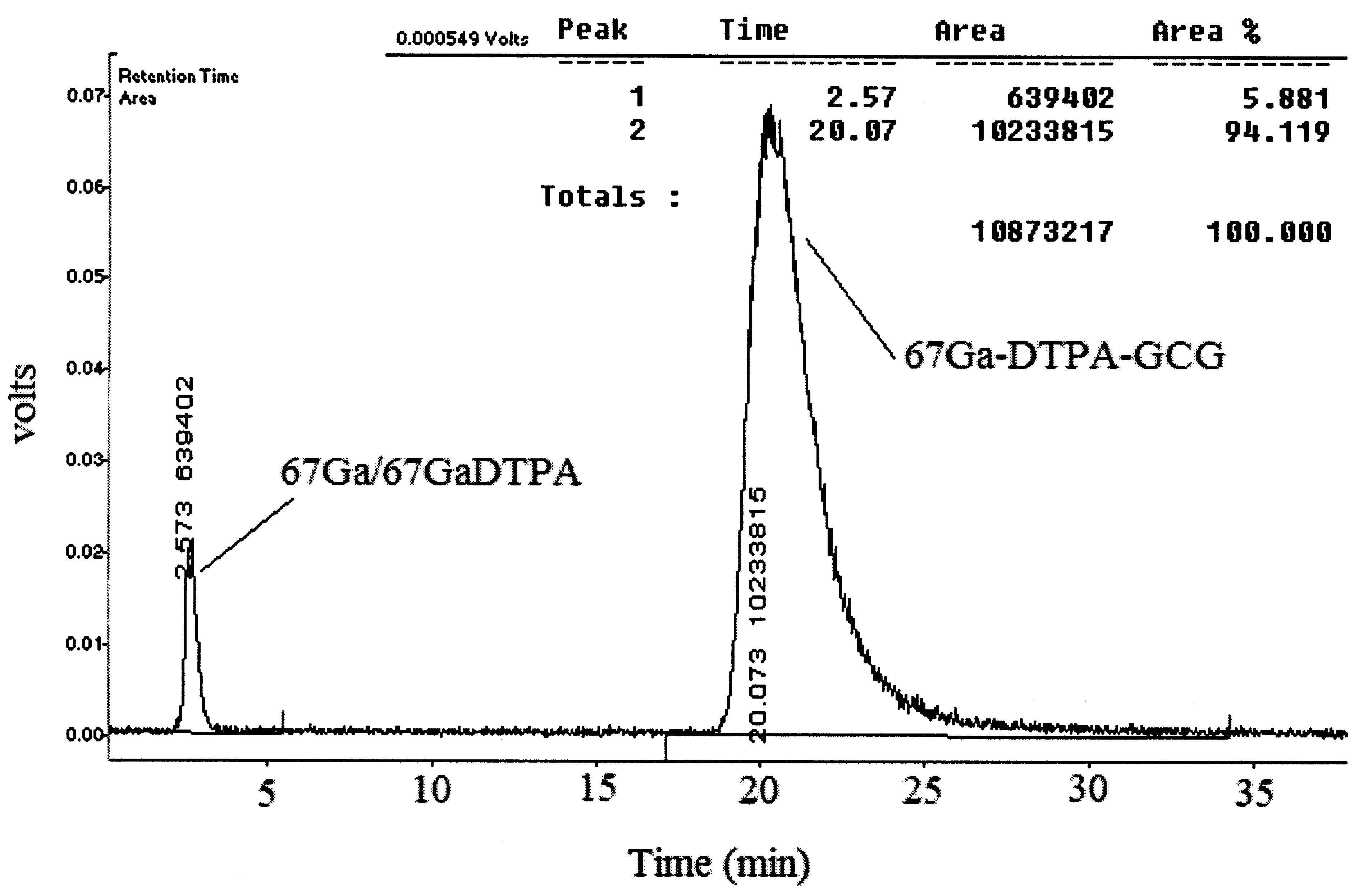
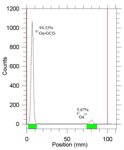
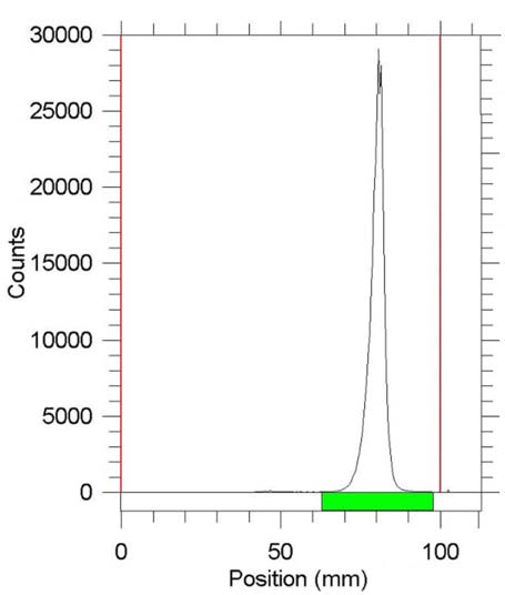 Fig. 2. ITLC of free 67Ga used in the radiolabeling (right) and 67Ga-GCG solution (left) on Whatman paper using 10 mM DTPA