Anterior chamber flare after femtosecond laser-assisted cataract surgery

Robin G. Abell, MB BS, Penelope L. Allen, PhD, Brendan J. Vote, FRANZCO PURPOSE: To determine whether postoperative ocular inflammation is less after femtosecondlaser–assisted cataract surgery than after conventional phacoemulsification (manual) cataractsurgery.
SETTING: Private clinic, Launceston, Tasmania, Australia.
DESIGN: Prospective consecutive investigator-masked nonrandomized parallel cohort study.
METHODS: Consecutive cataract patients who had femtosecond laser–assisted cataract surgery ormanual cataract surgery by the same surgeon at a single center were assessed. The primaryendpoint was postoperative aqueous flare measured by laser flare photometry at 1 day and 4 weeks.
Secondary endpoints included retinal thickness measured by optical coherence tomography andslitlamp examination findings at 4 weeks.
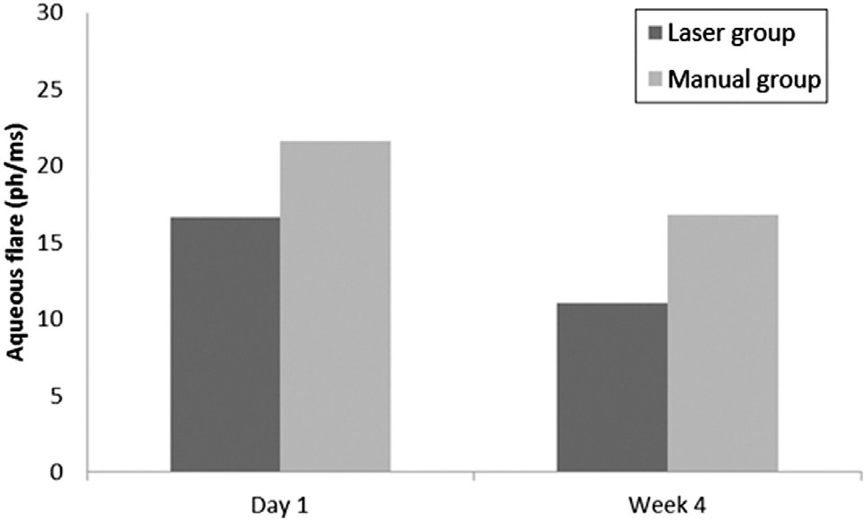
RESULTS: The per-protocol population comprised 176 patients (100 in laser group; 76 in manualgroup). Postoperative aqueous flare was significantly greater in the manual cataract surgery groupat 1 day (PZ.0089) and at 4 weeks (PZ.003). There was a significant correlation between effectivephacoemulsification time and 1-day postoperative aqueous flare (r Z 0.35, P<.0001). The increasein outer zone thickness measured by optical coherence tomography was less in the laser group(PZ.007).
CONCLUSION: Anterior segment inflammation was less after femtosecond laser–assisted cataractsurgery than after manual cataract surgery, and this appeared to be due to a reduction in phaco-emulsification energy.
Financial Disclosure: No author has a financial or proprietary interest in any material or methodmentioned.
J Cataract Refract Surg 2013; 39:1321–1326 Q 2013 ASCRS and ESCRS Advances in surgical equipment, ophthalmic viscosur- surgical trauma and the resulting inflammation. At gical devices, and phacoemulsification have led to a present, there is early evidence of a reduction in post- reduction in surgical trauma.The introduction of operative swelling at the macula with femtosecond femtosecond laser–assisted cataract surgery has led laser–assisted cataract surgery over manual cataract to further reductions in phacoemulsification energy re- quirements.Combined with automated corneal in- Postoperative inflammation is associated with a cisions and anterior capsulotomy, femtosecond breakdown of the blood–aqueous barrier as a result of laser–assisted cataract surgery may further reduce surgical trauma–induced prostaglandin production.The inflammation generally manifests as mild iritis,corneal edema, increased cells and protein (flare) inthe anterior chamber, and hyperalgesia or painEven in its mildest form, postoperative inflammation remains Final revision submitted: May 29, 2013.
a challenge, and patients have high expectations for rapid visual recovery and minimal associated pain.
From the Tasmanian Eye Institute (Abell, Allen, Vote) and the Laun- It is not known whether femtosecond laser–assisted ceston Eye Institute (Vote), Launceston, Tasmania, Australia.
cataract surgery lens fragmentation leads to an increase Corresponding author: Brendan J. Vote, FRANZCO, Launceston Eye in particulate matter in the anterior chamber, which Institute, 36 Thistle Street West, Launceston 7250, Australia.
may exacerbate inflammatioThis study was per- formed to compare postoperative inflammation using ANTERIOR CHAMBER FLARE AFTER LASER-ASSISTED CATARACT SURGERY anterior chamber flare between femtosecond laser–as- concentric regions were split into 4 zones each as follows: sisted cataract surgery and manual cataract surgery.
superior, temporal, inferior, and nasal. The mean of the4 values in each ring comprised the inner OCT measurementand outer OCT measurement, respectively. The inner ring and outer ring had a radius of 1.5 mm and 3.0 mm, Anterior chamber aqueous flare was measured objectively This was a prospective consecutive investigator-masked using laser flare photometry (Kowa FM-600). This instru- nonrandomized parallel cohort study performed at a single ment and its operation have been described in detail.
center. The study was approved by the Tasmanian Human Anterior chamber flare was measured by a masked investi- Research Ethics Committee and was performed in accor- gator who was unaware of the patient’s treatment group.
dance with the Declaration of Helsinki and its subsequent re- Measurements were taken within 1 week preoperatively as well as 1 day and 4 weeks postoperatively. Seven measure- Consecutive patients who were older than 18 years and ments were taken under scotopic conditions without phar- planned to have femtosecond laser–assisted cataract surgery macologic pupil dilation. The 2 extreme values were or manual cataract surgery with insertion of a posterior excluded from the mean value, according to the manufac- chamber intraocular lens (IOL) were enrolled in the study.
turer’s guidelines. Measurement conditions were kept All patients were given the option to have femtosecond consistent for all patients. The 5 measurements were aver- laser–assisted cataract surgery. Patients who elected to aged to a single score in photons per millisecond.
have femtosecond laser–assisted cataract surgery were Postoperatively, patients were seen at 1 day and 4 weeks.
placed in the laser group, and the remaining patients were Study assessment at the postoperative visits included slit- lamp examination, fluorescein staining for corneal epithelial Patients were excluded from the study if they had a preop- erosions, and intraocular pressure (IOP) measurement using erative flare of more than 15 photons per millisecond (ph/ Goldmann applanation tonometry. Dilated fundoscopy was ms) measured with a laser flare photometer without phar- performed at 4 weeks. Concomitant medications used to macologic pupil dilation, inflammatory or infectious pathol- treat inflammation related to cataract surgery were recorded ogy of the eye, history of postoperative intraocular infection at the preoperative and at the postoperative visits.
in the fellow eye, glaucoma, posttraumatic cataract, exfolia-tion syndrome, diabetic retinopathy, history of uveitis, and pathology requiring the use of topical or systemic antiinflam-matory or antiinfectious agents. Patients taking medications All patients instilled topical ketorolac and chloramphen- known to cause fluctuations or alterations in anterior cham- icol for 2 days before the procedure. On the day of surgery, ber protein composition or effect photometry flare values patients received topical anesthesia and pupil dilation with were excluded.Patients were also excluded from analysis a gel formulation consisting of phenylephrine 2.5%, cyclo- if they had an intraoperative complication of vitreous loss pentolate 1.0%, tropicamide 1.0%, lidocaine hydrochloride or complicated capsule rupture or had implantation of an jelly 2.0% (Xylocaine), and diclofenac 0.1%. The femtosecond laser procedure has been After the laser proce-dure, the patient was transferred to the operating room for regional anesthesia via sub-Tenon injection. Patients in themanual group also had regional anesthesia. The interval be- Eligible patients were included in the study after going tween the completion of the laser treatment and the initiation through an extensive preoperative assessment. Patients of operative cataract surgery was recorded for all patients in also had clinical and fundoscopic examinations using slit- lamp biomicroscopy. Evaluations included optical coherence Intraoperatively, corneal incisions were made manually tomography (OCT) (Zeiss Cirrus HD-OCT 4.0, Carl Zeiss using a 2.75 mm keratome and a 1.20 mm side-port blade.
Meditec AG), axial length (AL) and biometry (IOLMaster Patients having the laser procedure had the cut anterior 4, Carl Zeiss Meditec AG), and laser flare photometry capsule removed using a capsulorhexis forceps, after which (Kowa FM-600, Kowa Co., Ltd.). The cataract grade was as- hydrodissection was performed. Lens segmentation was sessed objectively using Scheimpflug imaging (Pentacam completed with the standard phacoemulsification procedure Nuclear Staging System, Oculus Optikger€ate (Megatron S4, Geuder AG). The effective phacoemulsifica- Optical coherence tomography measurements were per- tion time (EPT) was recorded for all patients. Patients who formed preoperatively within 2 weeks of the surgery and had manual cataract surgery had a continuous curvilinear postoperatively at 4 weeks. The same trained individual ob- capsulorhexis, hydrodissection, and phacoemulsification.
tained each scan and was masked to the patient’s treatment After successful removal of lens cortex, both cohorts had group. The repeatability and reproducibility of time-domain IOL placement in the capsular bag. All surgical characteris- tics except those related to the laser procedure were kept Macular measurements were taken after pupil dilation.
Scans were performed using a default AL (24.46 mm) and Postoperatively, all patients were prescribed topical chlor- refractive error to allow consistency with usual practice.
amphenicol, dexamethasone, and ketorolac 4 times a day for Scans were accepted if they were free of artifact. The instru- ment software automatically determined retinal thickness asthe distance between the internal limiting membrane and retinal pigment epithelium (RPE). Measurements were pro-vided for a central area as well as for 2 concentric regions.
The primary endpoint was aqueous flare measured with The central area (foveal region) had a radius of 0.5 mm (cen- laser flare photometry 1 day and 4 weeks after cataract sur- tral macular thickness [CMT]). The inner and outer gery. Secondary endpoints included EPT, fortified balanced J CATARACT REFRACT SURG - VOL 39, SEPTEMBER 2013 ANTERIOR CHAMBER FLARE AFTER LASER-ASSISTED CATARACT SURGERY salt solution (BSS Plus) fluid volume used during surgery, in-terval between the laser procedure and initiation of manualsurgery steps, postoperative IOP, and the change in retinalthickness from baseline measured by OCT. Retinal thicknessmeasured by OCT included central thickness and the meanof the 4 inner pericentral and 4 outer quadrants separately,as described above, at 4 weeks. An ophthalmologist assessedpatients at 1 day and 4 weeks using slitlamp biomicroscopy(anterior segment and fundus).
All data were imported into Stata 12 (Stata Corp LP) for analysis and examined with descriptive and frequency ana-lyses. Categorical data were analyzed using the chi-squaretest. Nonparametric continuous data were transformed to Figure 1. Mean aqueous flare in the laser group and manual group approximate a normal distribution, and t tests were used 1 day and 4 weeks after cataract surgery (ph/ms Z photons per to detect differences in means. Correlations between EPT and 1-day postoperative aqueous flare and between 1-dayaqueous flare and 4-week aqueous flare were assessed usingthe Pearson correlation coefficient (r). Regression models mean aqueous flare was 11.1 G 8.1 ph/ms and 14.6 with post-estimation diagnostics were run to evaluate the G 10.7 ph/ms, respectively (PZ.003) ). There relationship between EPT, balanced salt solution used, andaqueous flare at 1 day and 4 weeks. All tests were 2 sided, was no difference in IOP between groups at 1 day.
and a P value less than 0.05 was considered significant.
There was a significant correlation between the 1-day A post hoc power analysis found that the study had statis- and 4-week aqueous flare score (r Z 0.51, P!.0001).
tical power greater than the 0.80 level at a 0.05 to detect There was also a significant correlation between EPT a difference in mean aqueous flare between the laser group and 1-day postoperative aqueous flare (r Z 0.35, and manual group given the sample size in each group.
The multiple regression of EPT and balanced salt so- lution use as predictors of aqueous flare at 1 day was The study analyzed 100 eyes in the laser group and 76 significant (F3,101 Z 8.6, P!.05). For each 1-unit eyes in the manual group. All eyes completed the (1-second) increase in EPT, a 0.63 increase in aqueous study. There were 53 (53%) men in the laser group flare at 1 day would be expected. For each 1-unit and 33 (56%) in the manual group. The mean age (1 mL) increase in balanced salt solution, a 0.05 in- was 72.5 years G 10.5 (SD) (range 41 to 94 years).
crease in aqueous flare at 1 day would be expected.
There was no significant difference in age, refractive The regression of EPT and balanced salt solution as error, AL, anterior chamber depth, smoking status, predictors of aqueous flare at 1 month was also signif- preoperative IOP, nonocular medical history, or ocular The mean interval from completion of laser lens The mean preoperative aqueous flare was 5.3 G fragmentation to initiation of manual cataract surgery 3.1 ph/ms in the laser group and 5.3 G 3.4 ph/ms in in the laser group was 35 G 16 minutes. There was a the manual group; the difference was not statistically trend toward increased aqueous flare at 1 day with significant (PZ.96). The mean cataract grade was greater intervals between laser treatment and cataract 2.8 G 0.8 and 2.9 G 0.8, respectively; the difference surgery; however, it was not significant.
was not statistically significant (PZ.77).
At 4 weeks, the mean increase in OCT measure- There was a significant difference in mean EPT be- ments (CMT, inner zone, and outer zone) from base- tween groups (P!.0001). The laser group had a lower line were greater in the manual group ().
mean EPT (0.94 G 3.47 seconds) than the manual There was a significantly larger increase in the outer group (6.5 G 4.3 seconds). Similarly, the laser group zone internal limiting membrane and RPE thickness required less balanced salt solution volume during the manual steps of surgery than the manual group No statistically significant differences were found in (175.2 G 71.8 mL versus 195.1 G 76.5 mL), although the slitlamp examination and fundoscopy results be- tween the treatment groups at 1 day and 4 weeks.
(PZ.08). There were no intraoperative complications Furthermore, none of the study participants in either group had raised IOP postoperatively or required At 1 day, the mean aqueous flare was 16.6 G 8.9 ph/ concomitant medication to treat postsurgical inflam- ms in the laser group and 21.8 G 12.0 ph/ms in the mation. There were no adverse effects related to the use of topical medications in either group.
J CATARACT REFRACT SURG - VOL 39, SEPTEMBER 2013 ANTERIOR CHAMBER FLARE AFTER LASER-ASSISTED CATARACT SURGERY Our original assumption was that residual lens ma- Table 1. Between-group comparison of the increase in OCT terial and particulate matter may be a consequence of measurements from baseline to 4 weeks.
femtosecond laser lens fragmentation, resulting in a higher incidence of postoperative inflammation as measured by aqueous flare. The interval between the completion of laser lens fragmentation and the initia- tion of manual cataract surgery may also have an effect on postoperative inflammation. Residual lens material after cataract surgery or traumatic rupture of the lens capsule is known to provoke intraocular inflamma- tionhowever, this does not appear to be the case in our study. The difference in aqueous flare readings between groups was most likely due to the reduction in EPT in the laser group. We also found no significant association between the interval and aqueous flare values, although generally the delay was less than45 minutes. Therefore, we believe lens particulatematter produced by the femtosecond laser during lens fragmentation has a minimal impact with short Several studieshave established the safety and intervals. The maximum permitted interval between efficacy of femtosecond laser–assisted cataract sur- laser lens fragmentation and manual surgery to gery; however, little is known about the postopera- limit postoperative inflammation must still be tive inflammation caused by surgical trauma and fragmented lens matter. The objective of our study In terms of secondary endpoints, there was no sig- was to compare postoperative inflammation after un- nificant between-group difference in CMT and inner eventful femtosecond laser–assisted cataract surgery zone change in retinal thickness from baseline with that after uneventful manual cataract surgery measured by OCT. The laser group had a significantly to assess the degree of surgical trauma. Laser flare lower increase in the outer zone retinal thickness on meter measurements showed that femtosecond OCT than the manual group. This is in agreement laser–assisted cataract surgery resulted in less with previous stAn association has been re- aqueous flare than manual cataract surgery at 1 day ported between phacoemulsification and the forma- and 4 weeks. The laser flare meter was used as an tion of significant clinical cystoid macular edema objective assessment of flare; its use for this purpose (CME).Most subclinical postoperative increases has been validated in terms of quantification, sensi- in retinal thickness are asymptomatic. Although this tivity, reproducibility, and reliabilitSlitlamp may represent the process that in its most advanced examination or other scoring methods may be less stages leads to the formation of it is unlikely sensitive, more prone to observer bias or error, and to be of clinical relevance unless symptomatic. Based on our results, femtosecond laser–assisted cataract Multiple factors can affect laser flare photometry surgery may offer advantages in controlling the phys- values.These include mydriatic agents, pupil size, iologic changes contributing to CME. However, the age, cataract, time of day, protein composition, and pa- clinical relevance of this is unknown. Randomized tient medicationTo reduce the probability of controlled trials have shown that clinical CME and laser photometric measurements being influenced by perifoveal thickening on OCT are largely prevented these factors, the factors were kept constant between by nonsteroidal antiinflammatory drugs (NSAIDs) patients and were taken into account when comparing thus, the additional benefit of the femtosecond laser values between patients and between serial measure- is unlikely to be of clinical significance. It is difficult ments over time. There was no difference in mean to know whether, independently, the femtosecond age between the groups. Multiple readings were taken laser would offer sufficient protection against CME to prevent sampling error. Medications that alter flare were assessed preoperatively, and patients were Limitations of this study include the use of topical excluded if they were taking any of these medications.
corticosteroids and NSAIDs during the perioperative All patients were assessed at roughly the same time of period. Many cataract surgeons treat inflammation day (early morning) and did not have dilating drops prophylactically using topical corticosteroid and before the test, and lighting conditions remained the nonsteroidal antiinflammatory medications.Both medications have been shown to be effective in the J CATARACT REFRACT SURG - VOL 39, SEPTEMBER 2013 ANTERIOR CHAMBER FLARE AFTER LASER-ASSISTED CATARACT SURGERY painhowever, corticosteroids carry the risk for adverse effects, such as increased IOP and delayed corneal healingThere were no unwanted effects related to the use of these topical medications in either group, and there was no difference in the 1-day IOP between groups. Although both groups were treated with the same drugs with the same frequency and duration, we cannot rule out differences in compliance between groups, which may have produced con- In conclusion, femtosecond laser pretreatment in cataract surgery significantly reduced the EPT. This appears to result in reduced postoperative ocular inflammation measured by aqueous flare and subse- quently a lower risk for macular edema. It is unknown whether this is solely due to the reduction in EPT aes FP, Costa EF, Cariello AJ, Rodrigues EB, Hofling- Lima AL. Comparative analysis of the nuclear lens opalescence between groups or is due to other processes.
by the Lens Opacities Classification System III with nuclear den-sity values provided by Oculus Pentacam: a cross-section studyusing Pentacam Nucleus Staging software. Arq Bras Oftalmol  Laser cataract surgery is safe and results in less postop- erative macular thickening than manual cataract surgery.
14. Paunescu LA, Schuman JS, Price LL, Stark PC, Beaton S, Ishikawa H, Wollstein G, Fujimoto JG. Reproducibility of nervefiber thickness, macular thickness, and optic nerve head mea-surements using StratusOCT. Invest Ophthalmol Vis Sci 2004;  There was less postoperative inflammation measured by aqueous flare and macular thickening after femtosecond 15. Leung CK, Cheung CY, Weinreb RN, Lee G, Lin D, Pang CP, Lam DSC. Comparison of macular thickness measurements be- tween time domain and spectral domain optical coherence to-  These results support the view that femtosecond laser– mography. Invest Ophthalmol Vis Sci 2008; 49:4893–4897.
Available at: .
assisted cataract surgery may be less traumatic than con- 22. Shah SM, Spalton DJ, Smith SE. Measurement of aqueous cells and flare in normal eyes. Br J Ophthalmol 1991; 75:348–352.
23. Biro Z, Balla Z, Kovacs B. Change of foveal and perifoveal thick- ness measured by OCT after phacoemulsification and IOL J CATARACT REFRACT SURG - VOL 39, SEPTEMBER 2013 ANTERIOR CHAMBER FLARE AFTER LASER-ASSISTED CATARACT SURGERY implantation. Eye 2008; 22:8–12. Available at: 28. Comstock TL, Paterno MR, Singh A, Erb T, Davis E. Safety and efficacy of loteprednol etabonate ophthalmic ointment 0.5% for the treatment of inflammation and pain following cataract sur- gery. Clin Ophthalmol 2011; 5:177–186. Available at: 26. Sivaprasad S, Bunce C, Wormald R. Non-steroidal anti- inflammatory agents for cystoid macular oedema following cata- ract surgery: a systematic review. Br J Ophthalmol 2005; J CATARACT REFRACT SURG - VOL 39, SEPTEMBER 2013

Source: http://images.ozongo.com/xpress/imex_corporativa/resources/image/Anterior_chamber_flare_after_femtosecond_laser_assisted_cataract_surgery_.pdf

Addiction research

ADDICTION RESEARCH: BELIEVING IN THE FUTURE1In global terms, alcohol research has been a growth industry in recent decades, as TomBabor has recently documented, and I believe the same could be documented for illicit drugs, forpsychopharmaceuticals, and for tobacco research. There is some reason to believe that the periodof growth may be coming to an end. The growth has, in any case, been highly
Microsoft word - newsletter issue 3 finalv2.doc

Ne wsletter “To facilitate & disseminate high quality homeopathy research.” Welcome to the third newsletter of the Homeopathy Research Institute, a newsletter for everyone who wants to understand and support scientific research into homeopathy. Homeopathy for women with menopausal symptoms: a review of the evidence Introduction All patients sought homeopat

Copyright © 2010-2014 Medical Pdf Finder