Do you want to buy antibiotics online without prescription? https://buyantibiotics24h.net/ - This is pharmacy online for you!
Severe n.pm
OPHTHALMIC OPHTHALMIC FORUM A relentless peripheral corneal ulcer Comments by: Chi-Cheong Wong, FRCS, FHKAM (Ophth), Jack A. Singer, MD, Peter G. Watson, FRCS, FRCOphth Case history
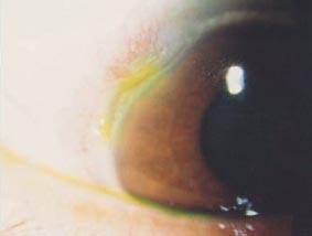
A 64-year-old lady with good past health first presented 7 years previously with left eye limbal congestion and a small peripheral corneal guttering ulcer at the nasal region (Figure 1) associated with mild pain. The ulcer healed with topical antibiotics and steroid in about 3 weeks.
A few months later, repeated ulceration developed which in-creased in size from 1 to 3 clock hours. Medicationsincluding topical steroid, antibiotics, timolol, and lubricants,were given. The patient was referred to a rheumatologist forsystemic examination. There were no signs or symptomssuggestive of an autoimmune disease and all the investigations,including immune markers, were negative. Systemic and
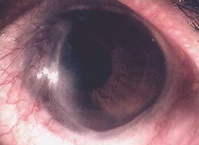
Figure 2. Healed ulcer with ingrowth of fibrovascular tissue.
subconjunctival steroids together with oral non-steroidal anti-inflammatory drugs were given because of the worsening
bandage contact lens were applied to seal the leakage in
condition with circumferential extension of the ulcer. A ban-
addition to topical steroid and antibiotic eyedrops. The
dage contact lens was also applied to promote healing. The
leaking gradually stopped with residual thinning and slight
ulcer gradually healed with ingrowth of fibrovascular tissue
ectasia. However, the leakage recurred 6 months later and
(Figure 2). The visual acuity in the affected eye was 0.3 unaided.
the procedure was repeated to treat the leakage. Topical
The systemic medications were gradually tapered and the
cyclosporin was given in addition to the other medications.
condition was maintained with low-dose topical steroid and
The perforation finally sealed with thinning and vasculariza-
antibiotics. The intraocular pressure was normal.
tion of the peripheral corneal gutter which extended for 270°from 3 to 12 o’clock. The visual acuity in the affected eye
One year later, thinning of the cornea and leaking of aqueous
was 3/60 and 0.8 in the other eye. All the topical medications
were noted central to the fibrovascular ingrowth at 11 o’clock.
Intraocular pressure was 10 mm Hg. Cyanoacrylate glue and
Several months later, corneal edema was noted and a cata- ract began to form (Figure 3). Visual acuity later decreased to hand movement. Endothelial count was approximately 1200 cell/mm2. The cornea was very thin at the inferonasal quadrant. Her right eye remained healthy apart from some crystalline deposits at the inferior limbus.
What are the comments on the pathogenesis, and previousand present management? Would you advise performingcataract surgery and corneal grafting for this patient?
Comments by Dr Chi-Cheong Wong, Senior Medical Officer, Tung Wah Eastern Hospital, Hong Kong, China Figure 1. Limbal congestion and peripheral corneal guttering
This 64-year-old lady suffered severe peripheral corneal
of the left eye.
disease with relentless progression and secondary
HKJOphthalmol Vol.5 No.1 OPHTHALMIC FORUM
Although quite controversial and with reports of manysevere complications, large corneal transplant has beenused to deal with this problem.2 Corneoscleral transplant-ation has also been advocated for the management of thistype of case, but complications such as glaucoma, epithelialdefect, rejection, and recurrence of disease have beenreported.3 The need for life-long immunosuppressionalso hinders the use of this operation. With the advancementof technology, artificial cornea is being developed withmore biocompatible material, which may help to managethese cases in the future.
After any surgical intervention, it is not the end but thebeginning of another challenging management problem. Frequent follow-up with special attention to any flare-up of
Figure 3. Corneal edema with cataract.
the disease and adequate and timely immunosuppressionis the key to success. Treatment of any complications such
complications of cataract and corneal decompensation. This
is a very challenging anterior segment problem, which isvery difficult to manage and should be comanaged by a
In summary, this is a very challenging anterior segment case.
corneal specialist, cataract surgeon, and physician.
The goal of management should be realistic and patientsshould be well informed about the prognosis, implications
For the initial management, I think a more aggressive
of surgical treatment, and side effects of toxic immuno-
approach should be taken after reviewing the history.
modifying agents. The ophthalmologist should act promptly
This idiopathic unilateral ulcerative keratitis is very rare and
and aggressively early in the course of the disease and
has a devastating visual outcome. Initial aggressive therapy
collaborate with a corneal specialist, cataract specialist, and
such as conjunctival resection, tissue adhesive and soft
physician to ascertain the best solution for the patient. With
contact lens application, systemic tetracycline for its anti-
advances in medicine such as potent immunosuppressive
collagenolytic effect, and topical medroxyprogesterone might
agents with fewer side effects and newer materials to make
have been used to retard the corneal destructive process.
artificial corneas, we hope that the discouraging picture of
Newer agents such as topical cyclosporin have been claimed
to be effective to halt the progression. Although the use oftopical FK 506 is still in clinical trials, it may be proved to be
References
a promising agent it the future as it is said to be 10- to 100-fold more potent than cyclosporin. However even with these
Riedel T, Seitz B, Langenbucher A, Naumann GO. Mor-phological results after eccentric perforating keratoplasty.:
modalities of treatment in the early phase, the disease may
progress and the search for any other underlying autoimmune
Watson PG, Richardson E. Large corneal transplants in
disease should be emphasized. Although more justified in
corneal destructive disease Klin Monatsbl Augenheilkd
bilateral cases, the use of systemic immunotherapy agents
such as methrotrexate, azathoprine, and cyclophosphamide
Hirst LW, Lee GA. Corneoscleral transplantation forend stage corneal disease. Br J Ophthalmol 1998;82:
can be considered for severe and recurrent attacks.
For the current management, this is a very difficult situ-ation. First of all, it is a very advanced peripheral corneal
Comments
thinning with 270° involvement, and any lamellar
by Dr Jack A. Singer, Associate Professor of Surgery
tectonic graft will be very difficult to perform surgically
(Ophthalmology), Dartmouth Medical School,
and adequate immunosuppression before the operation is
Dartmouth-Hitchcock Clinic, 40 South Main Street,
very important as there is a high chance of recurrence. Also
Randolph, VT 05060, USA
eccentric corneal graft may be served for tectonic purposein the initial phase of treatment.1 Definitive penetrating
This lady has a chronic progressive peripheral ulcerative
keratoplasty can be done at a later stage after the tectonic
keratitis, known as Mooren's ulcer, which is now compli-
graft. The cataract can be managed during the keratoplasty
cated by corneal decompensation and cataract. As this is
in an open sky manner but special attention to technique is
an autoimmune disease, the use of topical and systemic
very important to avoid excessive postoperative inflam-
cyclosporin has been recommended in light of its ability to
mation. Management of any coexisting anterior segment
increase the population of suppressor T cells.
problems should be kept in mind. In the bag placement ofintraocular lens, meticulous cleaning of the cortex, and use
Attempts at reparative corneal surgery are seldom success-
of heparin-coated intraocular lenses are measures that can
ful unless the underlying Mooren's disease activity has been
add to the reduction of postoperative inflammation.
controlled. Even when the disease has ‘burned itself out,’
HKJOphthalmol Vol.5 No.1 OPHTHALMIC FORUM
attempts at corneal grafting are usually associated with
starting about 2 mm from the limbus. The ulcer advances
recurrence of the Mooren's ulcer and destruction of the
both circumferentially and centrally, preceeded by thicken-
graft. Wide conjuctival resection to bare sclera, followed by
ing and edema of the cornea. The ulcer usually has an
resection of the overhanging lip of the ulcerating cornea
undermined edge but this is not a feature in this case.
and application of tissue adhesive will presumably removethe source of the antibody and inflammatory cells that
As the ulcer advances centrally, the endothelium becomes
may be mediating the ulcerative reaction. Additionally,
affected and eventually corneal decompensation occurs.
amniotic membrane transplantation may promote healing
Perforation, if it occurs, is usually the result of minor trauma.
of the damaged and thinned corneal tissue.
Cataract is a late complication. There is never any accom-panying systemic disease nor any associated scleritis. The
If amniotic membrane transplantation does not result in
limbus is congested. Full human leukocute/lymphocyte
sufficient healing and regeneration of peripheral corneal tissue,
antigen testing may confirm the diagnosis. Helminthic
then lamellar tectonic grafting would be needed prior to de-
infestations should be excluded or treated. Mooren’s ulcer
finitive central penetrating grafting. Immunosuppression
is often bilateral and any minor truama to the other eye could
therapy prior to corneal grafting may help prevent a recur-
easily result in the process starting there.
rence or rejection. An ‘open sky’ extracapsular cataract ex-traction with posterior chamber intraocular lens implantation
Treatment is unsatisfactory and rarely curative. In Aravind
can be performed together with the penetrating keratoplasty.
in India, where this disease is common, some success hasbeen achieved by the use of cyclosporin eye ointment. Cor-
Comments
neal transplantation is rarely successful as the ulcer recurs
by Dr Peter G. Watson, 17 Adams Road, Cambridge CB3
in the graft. If cataract surgery or transplantation is perform-
9AD, UK
ed, this needs to be accompanied by full immunosuppres-sion as the evidence is that the ulcer is an immune response
This patient has the typical history and appearance of a
to calgranulin C which is uniquely present in the corneal
Mooren’s ulcer. Pain is usually a marked feature of this
stroma. This explains why the epithelium and endothelium
disease although it is not necessarily severe. The character-
remain unaffected until very late in the disease. Cataract
istic features are a painful peripheral corneal ulceration
extraction must be done through a scleral tunnel. Complimentary Copy
A complimentary copy of the journal can be obtained from the College ofOphthalmologists of Hong Kong on request. The College of Ophthalmologists of Hong Kong EGE OFOPHTHALMOLOGISTS Room 802, 8/F, Hong Kong Academy of Medicine Jockey Club Building 99 Wong Chuk Hang Road, Aberdeen Hong Kong, China. Tel: (852) 2761 9128 Fax: (852) 2715 0089 E-mail: cohk@netvigator.com HKJOphthalmol Vol.5 No.1
The notion of cultural history and cultural studies in general, as usually employedin contemporary academic discourse, is derived from social anthropology. The culturalhistorian reads texts and other historical sources and studies artifacts, not so much asdiscursive expositions. Rather, like an anthropologist studying live behavior, thehistorian seeks both to discover the ways people in the socie
Term Effective: Full Title: Advanced Cardiac Life Support (limit to 50 characters including spaces) If this is a variable unit course, then the relationship between units and any difference in expected SLO’s should be explained. Student Learning Outcomes: (Enter the SLO’s in an outline format. Use the Ctrl + Tab keys to indent for subtopics.) At the conclusion of this cour
 OPHTHALMIC
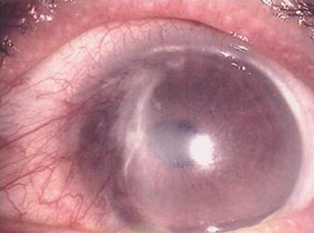 OPHTHALMIC FORUM